
Page 10 - Read Online
P. 10
Page 4 of 12 Bryan et al. Plast Aesthet Res 2022;9:53 https://dx.doi.org/10.20517/2347-9264.2022.39
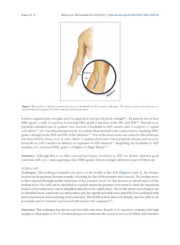
Figure 1. Illustration of relevant anatomy for nerve to brachialis to AIN transfer technique. The donor nerve to the brachialis is
transferred to the recipient AIN. AIN: Anterior interosseous nerve.
[21]
transfers regained grip strength, and 3/4 regained at least partial pinch strength . All patients met at least
MRC grade 3, with 3/4 patients recovering MRC grade 4 function of the FPL and FDP . Hawasli et al.
[21]
reported a detailed case of a patient who received a brachialis to AIN transfer after a complete C7 spinal
cord injury . At 3 months postoperatively, the patient demonstrated early reinnervation, regaining MRC
[18]
grade 3 strength in the FDP and FPL of the left hand . One of the most recent case series for this technique
[18]
was described by Souza et al. in 2020, where 11 patients had lower brachial plexus injuries and received
brachialis to AIN transfers in addition to supinator to PIN transfers . Regarding the brachialis to AIN
[22]
transfers, 8/11 recovered MRC grade 3 or higher on finger flexion .
[22]
Summary: Although this is an older reported technique, brachialis to AIN has shown relatively good
outcomes, with 14/17 cases regaining at least MRC grade 3 flexion strength within two years of follow-up.
ECRB to AIN
Technique: This technique transfers the nerve to the ECRB to the AIN [Figures 2 and 3]. An oblique
incision in the proximal forearm is made, following the line of the pronator teres muscle. The median nerve
is then exposed through medial retraction of the pronator teres. On the anterior or lateral aspect of the
median nerve, the AIN can be identified as it passes under the pronator teres muscle, while the superficial
branch of the radial nerve can be identified adjacent to the radial artery. The ECRB motor nerve branch can
be identified most commonly as a trifurcation with the superficial radial nerve and PIN. It is confirmed with
nerve stimulation demonstrating wrist extension. The ECRB is then dissected distally, and the AIN is cut
proximally and the transfer is performed with tension-free coaptation .
[23]
Outcomes: This technique has shown very favorable outcomes. Bertelli et al. reported 4 patients with high
median or ulnar palsy or C7-T1 brachial plexus root avulsions who received nerve to ECRB to AIN transfers