
Page 44 - Read Online
P. 44
Dellon. Plast Aesthet Res 2022;9:45 https://dx.doi.org/10.20517/2347-9264.2022.13 Page 7 of 15
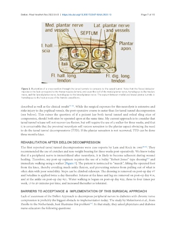
Figure 3. Illustration of a cross-section through the tarsal tunnels to compare to the carpal tunnel. Note that the flexor/abductor
muscles in the foot correspond to the thenar muscles in hand, and cover the roof of the medial plantar nerve, homologous to the median
nerve, and the lateral plantar nerve, homologous to the lateral plantar nerve. The septum between medial and lateral plantar tunnels is
homologous to the hook process of the hamate carpal bone.
described as well as the clinical results [78-80] . While the surgical exposure for this neurolysis is extensive and
risks injury to the popliteal vessels, the post-operative course is easier than for tarsal tunnel decompression
(see below). This raises the question of if a patient has both tarsal tunnel and soleal sling sites of
compression, should both sites be operated upon at the same time. My current approach is to consider that
tarsal tunnel release will not recover toe flexion, but will require the use of a walker for three weeks, and that
it is conceivable that the proximal neurolysis will restore sensation to the plantar aspect obviating the need
to do the tarsal tunnel decompressions (TTD). If the plantar sensation is not recovered, TTD can be done
three months later.
REHABILITATION AFTER DELLON DECOMPRESSION
The first reported tarsal tunnel decompressions were case reports by Lam and Keck in 1962 [80,81] . They
recommended the use of crutches and non-weight bearing for three weeks post-operatively. We know today
that if a peripheral nerve is immobilized after neurolysis, it is likely to become adherent during wound
healing. Therefore, my post-op regimen requires the use of a bulky “Robert Jones” type dressing and
[82]
immediate walking using a walker [Figure 7]. The patient is instructed to “march”, lifting the operated foot
from the knee, thereby avoiding much ankle flexion, and preventing sutures from pulling out of what is
often skin with poor sensibility. Steps can be climbed sideways. The dressing is removed on post-op day #7
and betadine is applied twice a day thereafter. Sutures at the knee and leg are removed on post-op day #14,
and at the ankle on post-op day #21. Water walking is begun on post-op day #22, three to four times per
week, 15 to 20 minutes per time, and increased thereafter as tolerated.
BARRIERS TO ACCEPTANCE & IMPLEMENTATION OF THIS SURGICAL APPROACH
Lack of awareness of the Dellon Approach to decompress peripheral nerves in diabetics with chronic nerve
compression is probably the biggest obstacle to implementation today. The study by Melenhorst et al., from
[31]
Zwolle in the Netherlands, best illustrates this problem . In that study, they asked physicians and diabetes
nurse educators the following questions: