
Page 30 - Read Online
P. 30
Amin. Plast Aesthet Res 2022;9:24 https://dx.doi.org/10.20517/2347-9264.2021.119 Page 5 of 10
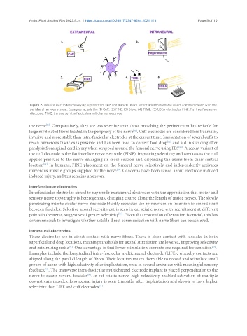
Figure 2. Despite electrodes conveying signals from skin and muscle, more recent advances enable direct communication with the
peripheral nervous system. Examples include the (1) Cuff; (2) FINE; (3) Sieve; (4) TIME; (5) USEA electrodes. FINE: Flat interface nerve
electrode; TIME: transverse intra-fascicular multichannel electrode.
the nerve . Comparatively, they are less selective than those breaching the perineurium but reliable for
[50]
large myelinated fibres located in the periphery of the nerve . Cuff electrodes are considered less traumatic,
[51]
invasive and more stable than intra-fascicular electrodes at the current time. Implantation of several cuffs to
reach numerous fascicles is possible and has been used to correct foot drop and aid in standing after
[52]
paralysis from spinal cord injury when wrapped around the femoral nerve using FES . A recent variant of
[53]
the cuff electrode is the flat interface nerve electrode (FINE), improving selectivity and contacts as the cuff
applies pressure to the nerve enlarging its cross-section and displacing the axons from their central
[54]
location . In humans, FINE placement on the femoral nerve selectively and independently activates
numerous muscle groups supplied by the nerve . Concerns have been raised about electrode induced
[55]
induced injury, and this remains unknown.
Interfascicular electrodes
Interfascicular electrodes aimed to supersede extraneural electrodes with the appreciation that motor and
sensory nerve topography is heterogenous, changing course along the length of major nerves. The slowly
penetrating interfascicular nerve electrode bluntly separates the epineurium on insertion to embed itself
between fascicles. Selective axonal recruitment is seen in cat sciatic nerve with recruitment at different
[56]
points in the nerve, suggestive of greater selectivity . Given that restoration of sensation is crucial, this has
driven research to investigate whether a stable direct communication with nerve fibers can be achieved.
Intraneural electrodes
These electrodes are in direct contact with nerve fibres. There is close contact with fascicles in both
superficial and deep locations, meaning thresholds for axonal stimulation are lowered, improving selectivity
[57]
[51]
and minimising noise . One advantage is that lower stimulation currents are required for sensation .
Examples include the longitudinal intra-fascicular multichannel electrode (LIFE), whereby contacts are
aligned along the parallel length of fibres. Their location makes them able to record and stimulate small
groups of axons with high selectivity after implantation, seen in several amputees with meaningful sensory
feedback . The transverse intra-fascicular multichannel electrode implant is placed perpendicular to the
[58]
nerve to access several fascicles . In rat sciatic nerve, high selectively enabled activation of multiple
[59]
downstream muscles. Less axonal injury is seen 2 months after implantation and shown to have higher
selectivity than LIFE and cuff electrodes .
[51]