
Page 103 - Read Online
P. 103
Page 6 of 14 Evans et al. Plast Aesthet Res 2022;9:34 https://dx.doi.org/10.20517/2347-9264.2021.134
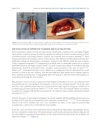
Figure 1. Vascularized bone flaps for lower extremity reconstruction of bony defects. (A) free fibula osteocutaneous flap. (B) in situ
medial femoral condyle flap isolated on descending geniculate vessels for the reconstruction of a small bony defect.
THE EVOLUTION OF OPERATIVE PLANNING AND FLAP SELECTION
Early reconstructive options in lower extremity trauma relied heavily on physical exam and duplex Doppler
ultrasound for surgical planning. Formal arteriography was utilized to evaluate vascular patency in lower
extremities injuries concerning ischemia, however, not routinely used for surgical planning [20,92] . Upon
surgical exploration, the traumatic nature of these injuries often distorts soft tissue planes and anatomic
landmarks, making the determination of adequate recipient vessels difficult within the zone of injury.
Today, computed tomography (CT) angiography is routinely used as a minimally invasive way to evaluate
distal limb perfusion and target recipient vessels . In early reconstructive efforts, it was believed that flap
[92]
anastomoses should be performed with a recipient vessel proximal to the zone of injury [3,28,32,92] .
Kolker et al. , determined that there was no difference in complication rates for flaps based on vessels
[31]
proximal, or distal to the zone of injury. With the knowledge that distal vessels can be adequate targets in
lower extremity reconstruction, CT angiography allows the surgeon to effectively look at distal patency in
vessels that pass through the zone of injury.
Flap selection in lower extremity reconstruction had changed considerably since 1854, when Hamilton first
described the use of a cross-leg flap for the treatment of chronic lower extremity wounds . In the late 1890s
[8]
and early 1900s, pedicled muscle flaps and vascularized bone flaps began to emerge as a useful tool in
reconstructing soft tissue and boney defects [85,93,94] . In the 1960s-1970s, microsurgical options in extremity
reconstruction emerged, with the first successful lower extremity free tissue transfer performed by Daniel
and Taylor in 1971 .
[95]
With the emergence of microsurgical techniques, free muscle transfers with skin grafting and myocutaneous
free flaps soon became the gold standard for lower extremity reconstruction [2,3,12-14,31,44,46,51] . Severe soft tissue
deficits of the lower extremity that were once considered too distal, or too large for pedicled flap
reconstruction, could now be covered with free tissue transfer.
In the late 1980s, the advent of the perforasome theory and perforator flaps further expanded flap options
for lower extremity reconstruction. New fasciocutaneous and muscle-sparing myocutaneous flaps proved
not only reliable in covering soft tissue deficits, but also demonstrated decreased donor site morbidity and
improved cosmesis [12,40,41,43] [Figure 2].