
Page 40 - Read Online
P. 40
cases is generally longer than the initial incision, both in with myotomy of the epitrochlear muscles using the
the proximal and distal directions. In the subcutaneous Z‑lengthening technique. It is of paramount importance
[16]
tissue, the identification of small regional sensory nerves that excision of the medial intermuscular septum and a
may be difficult because they are frequently incorporated complete opening of the distal septum between the FCU
in the scar tissue from previous surgeries. It is not and the flexor‑pronator muscle group are performed.
uncommon to find that one or more of these nerves have If the transposition has been accomplished properly,
been severed. Possible neuromas must be removed, [3,14,19] the nerve will lie in its new location without areas of
and proximal nerve stumps must be cauterized and compression or kinking; (2) the ulnar nerve was previously
positioned in good‑quality soft tissue, such as the triceps transposed anteriorly and superficially, but there is
muscle. [3,14,19] In cases in which the ulnar nerve is entrapped currently severe fibrosis that renders nerve debridement
in firm, fibrous scar tissue, it is advisable to begin the difficult. If the intermuscular septum was not released
exploration proximal to the region of the previous incision during the previous surgery, the nerve passes over the
to identify the nerve in a healthy area. Progressing distally, septum, which dislocates the nerve from beneath, creating
the nerve is then released from the scar. Depending compression. In other cases, the nerve may be found atop
on the technique used during the first surgery, the the epitrochlear bone as a consequence of an erroneous
following three different situations may be encountered: transposition or of a failure of the soft tissue anchorage.
(1) the nerve has been decompressed and is still in the This situation creates tension along the nerve, resulting
epitrochlear‑olecranon channel; (2) the nerve is outside of in acute angulation and kinking of the nerve at Osborne’s
the epitrochlear‑olecranon channel because dynamic nerve arcade or at the deep distal septum at the level of the
instability has occurred with recurrent anterior subluxation FCU. External neurolysis and submuscular transposition
during elbow flexion, or because it has been transposed are performed as described in section A [Figures 2‑5]; and
anteriorly in the subcutaneous tissue; or (3) the nerve is (3) the ulnar nerve was previously already transposed.
outside of the epitrochlear‑olecranon channel because it Surgery then commences with identification of the nerve
has been transposed anteriorly under the flexor‑pronator proximal and distal to the scarred area, isolation of the
muscles. Regardless of where the nerve is located, the nerve from the point of fibrosis up to the entrance in the
presence of scar tissue is a consistent pattern, which epitrochlear muscles, and decompression of the arcade
increases both the difficulty of the dissection and the risk of Struthers, the intermuscular septum proximally, and
of nerve damage. In these cases, identification of the nerve the deep flexor‑pronator septum distally. Release of the
distal to the cubital channel at the FCU muscle entrance nerve at the entrance, exits, and beneath the muscular
is recommended. From there, dissection proceeds in a channel is then performed. The nerve is generally found
distal to proximal direction. Once the nerve and potential
compression areas have been released, the following
different anatomical situations may be encountered:
(1) the ulnar nerve was previously decompressed only
and is still located in the epitrochlear‑olecranon channel.
Proceed with anterior submuscular transposition
a
a b
c d
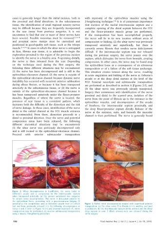
Figure 2: When decompression is insufficient, the nerve kinks at
Osborne’s arcade and is compressed by the intermuscular septum
when transposed anteriorly. (a) The patient underwent two surgeries
for simple nerve decompression. The nerve is dislocated anterior to b
the epitrochlear bone, presenting with a pseudoneuroma bulging (*)
proximal to the compression area at Osborne’s arcade level (>) which Figure 3: Failed nerve decompression treated with superficial anterior
had not been previously released (<); (b) following decompression at transposition. (a) The ulnar nerve (*) is fibrotic (<>), swollen, and hard
zones 4 and 5 (refer to Figure 1) (<) and external neurolysis (*), nerve to palpation; (b) the intermuscular septum (white arrows) and the distal
transposition may be performed; (c) anterior submuscular transposition deep septum in zone 5 (black arrows) were not released during the
using a muscle Z‑lengthening procedure initial surgery
180 Plast Aesthet Res || Vol 2 || Issue 4 || Jul 15, 2015