
Page 54 - Read Online
P. 54
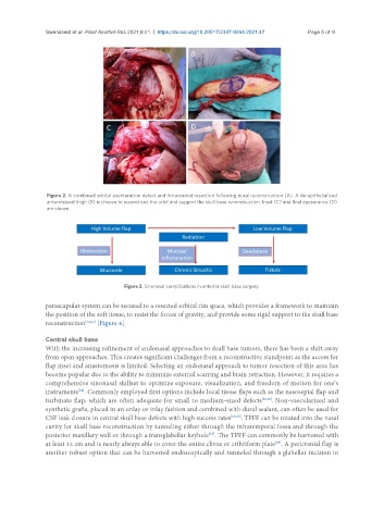
Swendseid et al. Plast Aesthet Res 2021;8:41  https://dx.doi.org/10.20517/2347-9264.2021.47  Page 5 of 9


                Figure 2. A combined orbital exenteration defect and intracranial resection following dural reconstruction (A). A de-epithelialized
                anterolateral thigh (B) is chosen to reconstruct the orbit and support the skull base reconstruction. Inset (C) and final appearance (D)
                are shown.


                                        Figure 3. Sinonasal complications in anterior skull base surgery.

               parascapular system can be secured to a resected orbital rim space, which provides a framework to maintain
               the position of the soft tissue, to resist the forces of gravity, and provide some rigid support to the skull base
               reconstruction [14,23]  [Figure 4].

               Central skull base
               With the increasing refinement of endonasal approaches to skull base tumors, there has been a shift away
               from open approaches. This creates significant challenges from a reconstructive standpoint as the access for
               flap inset and anastomosis is limited. Selecting an endonasal approach to tumor resection of this area has
               become popular due to the ability to minimize external scarring and brain retraction. However, it requires a
               comprehensive sinonasal skillset to optimize exposure, visualization, and freedom of motion for one’s
               instruments . Commonly employed first options include local tissue flaps such as the nasoseptal flap and
                         [24]
               turbinate flap, which are often adequate for small to medium-sized defects [25,26] . Non-vascularized and
               synthetic grafts, placed in an onlay or inlay fashion and combined with dural sealant, can often be used for
               CSF leak closure in central skull base defects with high success rates [27,28] . TPFF can be rotated into the nasal
               cavity for skull base reconstruction by tunneling either through the infratemporal fossa and through the
                                                                  [15]
               posterior maxillary wall or through a transglabellar keyhole . The TPFF can commonly be harvested with
               at least 12 cm and is nearly always able to cover the entire clivus or cribriform plate . A pericranial flap is
                                                                                      [29]
               another robust option that can be harvested endoscopically and tunneled through a glabellar incision to