
Page 64 - Read Online
P. 64
Apaydin. Plast Aesthet Res 2019;6:9 I http://dx.doi.org/10.20517/2347-9264.2018.73 Page 3 of 7
A B C
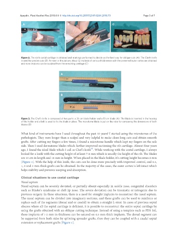
Figure 2. The sixth costal cartilage is obtained and markings performed to decide on the best way for oblique cuts (A); The Chef’s knife
is used for precise cuts (B); As seen in the picture, about 12 implants of various thicknesses with the preserved outer cortex are obtained
and more implants can be sculpted from the remaining cartilage (C)
A
B
Figure 3. The Chef’s knife is composed of two parts: a 26 cm blade holder and a 13 cm blade (A). The blade is inserted in the housing
of the holder and a bolt is used to fix the blade in place. The microtome blade is put on the ruler for comparing the dimensions of both
blades (B)
What kind of instruments have I used throughout the past 10 years? I started using the microtomes of the
pathologists. They were longer than a scalpel and very helpful to make clean long cuts and obtain smooth
grafts. After cutting my fingers a few times, I found a microtome handle which kept my fingers on the safe
side. Then I used dermatome blades which further improved sectioning the rib cartilage. Almost four years
[6]
ago, I found the ideal blade which I call as Chef’s knife . While working with the costal cartilage, I always
looked for a knife with the cutting height of at least 7-8 mm which is usually the height of the rib. The blades
are 13 cm in length and 14 mm in height. When placed in the blade holder, it’s cutting height becomes 8 mm
[Figure 3]. With the help of this knife, the cuts can be done more precisely with improved control, and 0.5,
1, 2 and 3 mm thick grafts can be obtained. In the majority of the cases, the outer cortex is left intact which
helps stability and prevents warping and absorption.
Clinical situations to use costal cartilage
Nasal septum
Nasal septum can be severely deviated, or partially absent especially in saddle nose, congenital disorders
such as Binder’s syndrome or cleft lip nose. The severe deviation can be traumatic or iatrogenic due to
previous surgery. In these situations, there is a need for straight implants to reconstruct the nasal septum.
The nasal septum can be divided into imaginary sections, and these grafts can be used to reinforce or
replace each of the segments (dorsal and or caudal) to obtain a straight L-strut. In cases of previous septal
abscess where all the septal cartilage is deficient, it is possible to reconstruct the entire septal cartilage by
using the grafts obtained with an oblique cutting technique. Instead of using a template such as PDS foil,
these implants of 1-2 mm in thickness can be sutured on 0.5 mm thick implants. The dorsal segment can
be supported from both sides by splinting spreader grafts, then they can be coupled with a caudal septal
extension or replacement grafts [Figure 4].