
Page 58 - Read Online
P. 58
Rajbhandari et al. Plast Aesthet Res 2019;6:8 I http://dx.doi.org/10.20517/2347-9264.2018.86 Page 7 of 10
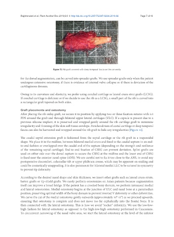
Figure 11. Rib graft covered with deep temporal fascia on the convexity
for the dorsal augmentation, can be carved into spreader grafts. We use spreader grafts only when the patient
undergoes extensive osteotomy, if there is evidence of internal valve collapse or if there is deviation of the
cartilaginous dorsum.
Owing to its curvature and elasticity, we prefer using conchal cartilage as lateral crura strut grafts (LCSG).
If conchal cartilage is deficient or if we decide to use the rib as a LCSG, a small part of the rib is carved into
a rectangular graft tapered on both sides.
Graft placements and osteotomy
After placing the rib onlay graft, we secure it in position by applying two or three fixation sutures with 5.0
PDS around the graft and through bilateral upper lateral cartilages (ULC). If a capsule is present due to a
previous silicone implant, it is preserved and wrapped gently around the rib cartilage graft to minimize
irregularity and thinning of the skin soft tissue envelope. Perichondrium of costal cartilage or deep temporal
fascia can also be harvested and wrapped around the rib graft to hide any irregularities [Figure 11].
The caudal septal extension graft is fashioned from the septal cartilage or the rib graft in a trapezoidal
shape. We place it in the midline, between bilateral medial crura and fixed to the caudal septum in an end-
to-end fashion or overlapped over the caudal end of the septum (depending on the strength and resilience
of the remaining septal cartilage). End-to-end fixation of CSEG can prevent deviation. Splint grafts are
used on either side over the dorsal septum to secure the CSEG at the midline and the lower end of CSEG
is fixed near the anterior nasal spine (ANS). We are careful not to fix it too close to the ANS, to avoid any
postoperative discomfort, columellar tilt or upper philtrum crease, which may be apparent on smiling and
could be cosmetically unappealing. It is also paramount for bilateral medial LLC to be sutured in symmetry
to prevent tip deformity.
According to the desired nasal shape and skin thickness, we insert other grafts such as lateral crura struts,
batten grafts or tip-shield grafts. We rarely perform osteotomies on Asian patients because augmentation
itself can improve a broad bridge. If the patient has a crooked bony dorsum, we perform intranasal medial
and lateral osteotomies. Medial osteotomy begins at the junction of ULC and nasal bone at a paramedian
position, preserving optimal width of the bony dorsum to prevent inverted V deformity or other deformities.
We curve the cut of the medial osteotomy gently outwards (approximately 10°-15°) as we proceed upwards,
ensuring that osteotomy is complete and does not move too far cephalically into the frontal bone. It is
then connected with the lateral osteotomy. This is how we avoid “rocker” deformity. We use the low-low-
high fashion for lateral osteotomy as opposed to the high-low-high osteotomy performed in Caucasians.
To circumvent narrowing of the nasal valve area, we start the lateral osteotomy at the level of the inferior