
Page 47 - Read Online
P. 47
Yi. Plast Aesthet Res 2019;6:7 I http://dx.doi.org/10.20517/2347-9264.2018.77 Page 3 of 7
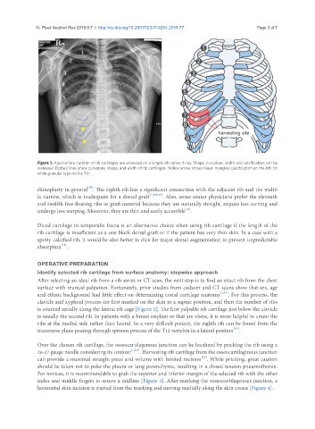
Figure 1. Appropriate number of rib cartilages are assessed on a simple rib series X-ray. Shape, curvature, width and calcification can be
reviewed. Dotted lines show curvature, shape, and width of rib cartilages. Yellow arrow shows linear marginal calcification on the 6th rib
while granular type on the 7th
[10]
rhinoplasty in general . The eighth rib has a significant connection with the adjacent rib and the width
is narrow, which is inadequate for a dorsal graft [1,6,8,11] . Also, some senior physicians prefer the eleventh
and twelfth free-floating ribs as graft material because they are naturally straight, require less carving and
[12]
undergo less warping. Moreover, they are thin and easily accessible .
Diced cartilage in temporalis fascia is an alternative choice when using rib cartilage if the length of the
rib cartilage is insufficient as a one block dorsal graft or if the patient has very thin skin. In a case with a
spotty calcified rib, it would be also better to dice for major dorsal augmentation to prevent unpredictable
[13]
absorption .
OPERATIVE PREPARATION
Identify selected rib cartilage from surface anatomy: stepwise approach
After selecting an ideal rib from a rib series or CT scan, the next step is to find an exact rib from the chest
surface with manual palpation. Fortunately, prior studies from cadaver and CT scans show that sex, age
and ethnic background had little effect on determining costal cartilage anatomy [1,11] . For this process, the
clavicle and xyphoid process are first marked on the skin in a supine position, and then the number of ribs
is counted serially along the lateral rib cage [Figure 2]. The first palpable rib cartilage just below the clavicle
is usually the second rib. In patients with a breast implant or that are obese, it is more helpful to count the
ribs at the medial side rather than lateral. In a very difficult patient, the eighth rib can be found from the
[11]
transverse plane passing through spinous process of the T12 vertebra in a lateral position .
Over the chosen rib cartilage, the osseocartilagenous junction can be localized by pricking the rib using a
26-27 gauge needle considering its contour [1,2,6] . Harvesting rib cartilage from the osseocartilaginous junction
[11]
can provide a maximal straight piece and volume with limited incision . While pricking, great caution
should be taken not to poke the pleura or lung parenchyma, resulting in a closed tension pneumothorax.
For novices, it is recommendable to grab the superior and inferior margin of the selected rib with the other
index and middle fingers to assure a midline [Figure 3]. After marking the osseocartilagenous junction, a
horizontal skin incision is started from the marking and moving medially along the skin crease [Figure 4].