
Page 22 - Read Online
P. 22
Page 4 of 15 Crowe et al. Plast Aesthet Res 2019;6:4 I http://dx.doi.org/10.20517/2347-9264.2018.70
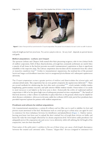
Figure 1. Carbon fiber partial foot prosthesis after Chopart amputation. The patient has remained active and is able run with this device
tubercle height partial foot prosthesis. The correct adaptive device - for any level - depends on patient factors
and goals.
Midfoot amputations - Lisfranc and Chopart
The eponyms Lisfranc and Chopart, both named after their pioneering surgeons, refer to two distinct levels
of midfoot amputation. Both of these disarticulations, although less commonly performed, are useful when
a paucity of soft tissue in the forefoot prevents successful transmetatarsal amputation or there is significant
instability at the respective joint. The Lisfranc amputation disarticulates all five metatarsals from the cuboid
[23]
or respective cuneiform bone . Similar to transmetatarsal amputations, the resultant loss of the peroneal
brevis and longus and dorsiflexor insertions leads to unopposed plantarflexion with subsequent equinovarus
deformity.
The Chopart amputation excises a greater portion of midfoot and disarticulates the talonavicular and
[24]
calcaneocuboid joints. This amputation has an even greater propensity for equinovarus deformity . There
are several methods for rebalancing the foot after such a procedure [25,26] , which include Achilles tendon
lengthening, gastrocnemius resection, and split anterior tibialis tendon transfer. Preservation of the ankle
in most instances is not helpful as the lever arm is short. Historically the utility of traditional midfoot
amputations is felt to be low given high rates of subsequent proximal revision amputations . The benefit of
[27]
this level, however, is that it allows the individual to place their foot on the ground, which may be helpful for
short distance ambulation. Advances in dynamic ankle foot orthosis (AFO) style partial foot prosthesis have
provided improved options for patients with midfoot amputations.
Prosthesis and orthosis for midfoot amputations
Like transmetatarsal amputations, a custom-fit orthosis and toe filler can be used to stabilize the foot and
prevent excess movement of the heel. Mechanisms such as a steel spring or carbon inlay can again be used
to counteract the force imbalance between the dorsiflexors and plantarflexors [Figure 1]. Patellar tendon-
bearing prostheses have been used to unload the short residual foot, although these devices are bulky and
lessen the value the extra length afforded by the chosen amputation level. Novel above-ankle prostheses that
adequately protect the amputation site and correct varus deformity, while still utilizing the full length of the
[27]
amputation, have also been described .
Preservation of the ankle joint in addition to the use of these inlays can cause a limb length discrepancy
between the treated and untreated sides. Thinner, “slipper-like” devices designed to minimize this