
Page 101 - Read Online
P. 101
Marsden et al. Plast Aesthet Res 2019;6:24 I http://dx.doi.org/10.20517/2347-9264.2019.14 Page 3 of 11
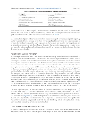
Table 1. Summary of the differences between nerve grafts and nerve transfers
Nerve graft Nerve transfer
2 Coaptations 1 Coaptation
~25% of available axons to target ~50% of available axons to target
Longer distance to target Shorter distance to target
Longer time to reinnervation Shorter time to reinnervation
Higher chance of motor end plate degeneration Allows for delayed reconstruction
Less specific Highly specific
Donor site morbidity Micro-neurolysis can preserve donor function
faster reinnervation to distal targets . Most commonly, this nerve transfer is used to restore motor
[22]
function, but it can be used to restore critical sensory function. The advantages of nerve transfer over nerve
grafts are well documented in the literature and are summarised in Table 1.
The combination of peripheral nerve reconstruction, such as nerve graft or transfer, along with importing
healthy vascularised tissue coverage in the form of free or pedicled tissue transfer is a useful technique to
optimise the local environment for nerve regeneration. Nerve grafts and transfers play a significant role
in extremity reconstruction and, depending on the defect characteristics (e.g., resection of major nerves
and muscular units), can be employed in combination with newer microsurgical techniques that have
developed in recent decades.
FUNCTIONING MUSCLE TRANSFER
Functioning muscle transfer (FMT) involves the transfer of a healthy donor muscle and its neurovascular
pedicle to a new location to assume a new function. Free functioning muscle transfer (FFMT) involves
restoring the circulation of the transferred muscle with microsurgical anastomosis to vessels at the recipient
site along with coaptation of the motor nerve. Pedicled innervated flaps maintain their vascular supply but
involve reorientation of the muscle and reinnervation from nerve transfer at the recipient site with a view to
altering the function of that muscle. Within several months, the transferred muscle becomes reinnervated
by the donor nerve, eventually begins to contract and ultimately gains independent function . FMT has
[23]
traditionally been limited to muscles with a single nerve for transfer (e.g., gracillis), because of the view
that segmental nerve supply would be an obstacle to reinnervation. However, we have previously published
a series of 11 functional quadriceps reconstructions using innervated rectus abdominis flaps, whereby
2-3 segmental nerves have been coaptated to cut femoral nerve branches at the recipient site. The overall
functional results were excellent with over 50% achieving M5 power [as per the Medical Research Council
[24]
(MRC) grading system] with a mean follow-up of 12 months and with minimal donor site complications .
The rectus abdominis has the added versatility of being used as a pedicled innervated flap for quadriceps
[25]
reconstruction, and its segmental innervation does not preclude its use as a functional muscle transfer .
The most reported FFMTs in the literature for STS extremity reconstruction are the gracillis [11,12,26] ,
[24]
latissimus dorsi (LD) [11,25,26] and rectus abdominis muscle [vertical (VRAM) or transverse (TRAM)] ,
with the other less commonly transferred muscles being the innervated medial gastrocnemius muscle
[27]
and rectus femoris [Figures 1-4]. FFMT can be performed in combination with nerve grafts and/or nerve
transfer, especially when the resection involves loss of major nerves and muscle units. Nerve grafts, either
standard or vascularised, are generally used with the aim of restoring critical sensory function, whereas
nerve transfer is primarily used to restore motor function.
LONG DONOR NERVE HARVEST WITH FFMT
In general, following sarcoma resection, there are usually native nerves available for coaptation to the
transferred muscle flap, which is advantageous as the length of nerve available with most flaps is short.