
Page 42 - Read Online
P. 42
Page 6 of 15 Ewing et al. Plast Aesthet Res 2024;11:22 https://dx.doi.org/10.20517/2347-9264.2024.11
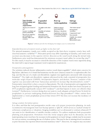
Figure 2. Technical pearls for the challenging microsurgery case in a radiated field.
Expanded dissection of recipient vessels: go higher on the chest wall
The internal mammary vessels, now widely accepted as the first-choice recipient vessels, have well-
described anatomic variability [48,49] . Bifurcation points may differ depending on laterality, and vessel cross-
sectional area decreases in the superior-inferior direction as the vessel travels along the sternal body.
[50]
Furthermore, prior work has demonstrated the minimum necessary vessel caliber for certain flap sizes .
For this reason, it may be necessary to extend the dissection of the recipient vessels more superiorly along
the chest wall to expose larger mammary vessel recipients for anastomoses.
Postoperative anticoagulation
The radiation-induced chronic inflammation renders vessels hypercoagulable , which raises concerns for
[8]
flap failure. Jakobsson et al. found that hematoma was the most common reason for re-exploration after free
flap, and that the use of a triple anti-thrombotic regimen was significantly associated with hematoma
formation . The triple anti-thrombotic regimen referenced in the study consisted of preoperative low
[51]
molecular weight heparin (LMWH), intraoperative heparin, and dextran. However, cessation of the
[51]
regimen was associated with fewer hematomas and did not increase flap thromboembolic events .
Postoperative immobility is often a concern in flap patients. The risk of postoperative anticoagulation has
long been mitigated against bleeding and hematoma formation risk. Recent studies have found that post-op
[45]
DVT prophylaxis significantly reduces DVT incidence , and that heparin is more cost-effective than
Lovenox . Furthermore, Lovenox dosing does not seem to reach adequate activated Factor Xa levels for
[52]
prophylaxis in patients undergoing head/neck/breast free flap procedures . Other centers have
[53]
implemented a 2-week prophylaxis protocol that lowers DVT but does not increase the incidence of
[54]
hematoma .
Salvage conduits: the bailout options
It is often said that the best postoperative results come with proper preoperative planning. As such,
reconstructive microsurgeons must have backup options: a “plan B”, which serves as a tactical remedy for
unforeseen complications or situations. Imaging modalities such as ultrasound or contrast-enhanced CT
can assist in the identification of suitable vascular conduits. The authors emphasize both preoperative
planning and preparation. Deviations from the initial surgical plan must be discussed, and the
team/operating room must have the necessary tools/equipment on hand. Salvage conduits may be necessary
for various reasons, necessitating the use of a bailout option. This may be due to missing/absent vasculature,
[55]
as prior literature has reported absent internal mammary veins . Moreover, the surgeon may encounter