
Page 35 - Read Online
P. 35
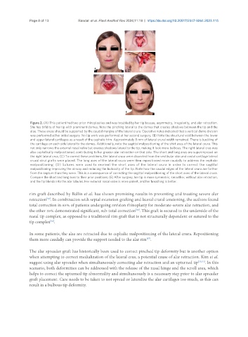
Page 8 of 13            Randall et al. Plast Aesthet Res 2024;11:18  https://dx.doi.org/10.20517/2347-9264.2023.115


                Figure 2. (A) This patient had two prior rhinoplasties and was troubled by her tip bossae, asymmetry, irregularity, and alar retraction.
                She has bifidity of her tip with prominent domes. Note the pinching lateral to the domes that creates shadows between the tip and the
                alae. These areas should be supported by the caudal margins of the lateral crura. Operative notes indicated that a vertical dome division
                was performed at her initial surgery. No tip work was performed at her second surgery; (B) Note the structural void between the lower
                and upper lateral cartilages as a result of the cephalic trim. Approximately 8 mm of lateral crural width remained. There is buckling of
                the cartilage on each side lateral to the domes. Additionally, note the sagittal malpositioning of the shirt axes of the lateral crura. This
                not only narrows the external nasal valve but creates shadows lateral to the tip, making it look more bulbous. The right lateral crus was
                also cephalically malpositioned, contributing to her greater alar retraction on that side. The short and long axes are superimposed on
                the right lateral crus; (C) To correct these problems, the lateral crura were dissected from the vestibular skin and costal cartilage lateral
                crural strut grafts were placed. The long axes of the lateral crura were then repositioned more caudally to address the cephalic
                malpositioning;  (D)  Sutures  were  used  to  reorient  the  short  axes  of  the  lateral  crura  in  order  to  correct  the  sagittal
                malpositioning-improving the airway and reducing the bulbosity of the tip. Note how the caudal edges of the lateral crura are farther
                from the septum than they were. This is a consequence of correcting the sagittal malpositioning of the short axes of the lateral crura.
                Compare the short and long axes to their prior positions; (E) After surgery, her tip is more symmetric, smoother, without alar retraction,
                and the tip blends into the alar lobules. Her external nasal valve is more patent, and her breathing is better.

               rim graft described by Ballin et al. has shown promising results in preventing and treating severe alar
                       [52]
               retraction . In combination with septal extension grafting and lateral crural tensioning, the authors found
               total correction in 65% of patients undergoing revision rhinoplasty for moderate-severe alar retraction, and
                                                                  [52]
               the other 35% demonstrated significant, sub-total correction . This graft is secured to the underside of the
               nasal tip complex, as opposed to a traditional rim graft that is not structurally dependent or sutured to the
               tip complex .
                         [52]
               In some patients, the alae are retracted due to cephalic malpositioning of the lateral crura. Repositioning
               them more caudally can provide the support needed to the alar rim .
                                                                       [37]
               The alar spreader graft has historically been used to correct pinched tip deformity but is another option
               when attempting to correct medialization of the lateral crus, a potential cause of alar retraction. Kim et al.
               suggest using alar spreader when simultaneously correcting alar retraction and an upturned tip [35,53] . In this
               scenario, both deformities can be addressed with the release of the nasal hinge and the scroll area, which
               helps to correct the upturned tip abnormality and simultaneously is a necessary step prior to alar spreader
               graft placement. Care needs to be taken to not spread or lateralize the alar cartilages too much, as this can
               result in a bulbous tip deformity.