
Page 46 - Read Online
P. 46
Chi et al. Plast Aesthet Res 2023;10:56 https://dx.doi.org/10.20517/2347-9264.2023.48 Page 3 of 13
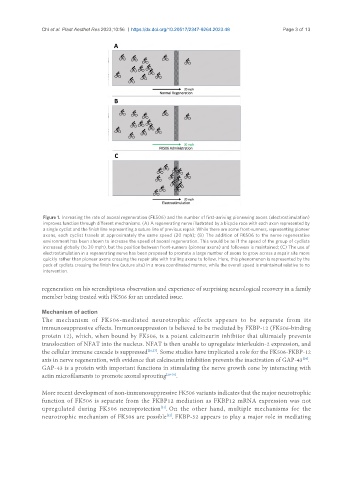
Figure 1. Increasing the rate of axonal regeneration (FK506) and the number of first-arriving pioneering axons (electrostimulation)
improves function through different mechanisms. (A) A regenerating nerve illustrated by a bicycle race with each axon represented by
a single cyclist and the finish line representing a suture line of previous repair. While there are some front-runners, representing pioneer
axons, each cyclist travels at approximately the same speed (20 mph); (B) The addition of FK506 to the nerve regenerative
environment has been shown to increase the speed of axonal regeneration. This would be as if the speed of the group of cyclists
increased globally (to 30 mph), but the position between front-runners (pioneer axons) and followers is maintained; (C) The use of
electrostimulation in a regenerating nerve has been proposed to promote a large number of axons to grow across a repair site more
quickly rather than pioneer axons crossing the repair site with trailing axons to follow. Here, this phenomenon is represented by the
pack of cyclists crossing the finish line (suture site) in a more coordinated manner, while the overall speed is maintained relative to no
intervention.
regeneration on his serendipitous observation and experience of surprising neurological recovery in a family
member being treated with FK506 for an unrelated issue.
Mechanism of action
The mechanism of FK506-mediated neurotrophic effects appears to be separate from its
immunosuppressive effects. Immunosuppression is believed to be mediated by FKBP-12 (FK506-binding
protein 12), which, when bound by FK506, is a potent calcineurin inhibitor that ultimately prevents
translocation of NFAT into the nucleus. NFAT is then unable to upregulate interleukin-2 expression, and
the cellular immune cascade is suppressed [26,27] . Some studies have implicated a role for the FK506-FKBP-12
axis in nerve regeneration, with evidence that calcineurin inhibition prevents the inactivation of GAP-43 .
[24]
GAP-43 is a protein with important functions in stimulating the nerve growth cone by interacting with
actin microfilaments to promote axonal sprouting [28-30] .
More recent development of non-immunosuppressive FK506 variants indicates that the major neurotrophic
function of FK506 is separate from the FKBP12 mediation as FKBP12 mRNA expression was not
upregulated during FK506 neuroprotection . On the other hand, multiple mechanisms for the
[31]
neurotrophic mechanism of FK506 are possible . FKBP-52 appears to play a major role in mediating
[32]