
Page 53 - Read Online
P. 53
Page 4 of 7 Sakai et al. Plast Aesthet Res 2023;10:45 https://dx.doi.org/10.20517/2347-9264.2023.18
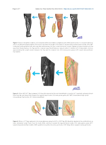
Figure 1. Based on lymphatic anatomy, ICG is injected at the most distal or peripheral parts of the lymphosomes in a recipient and donor
sites to visualize the lymph axialities. ICG is also injected at the border of the defect to visualize the proximal lymph axiality. After LIFT
is elevated and transferred with microvascular anastomoses, the flap is inset; the lymph vessels’ stumps are approximated with the
superficial fascia fixation, the flap and the recipient superficial fascia are sutured with 2-3 stitches of 3-0 absorbable stitches,
approximating the lymph vessels between the flap and the recipient site. ICG: indocyanine green; LIFT: lymph-interpositional-
flap transfer.
Figure 2. When ALT-LIFT flap is planned, ICG should be injected at the mid-lateral thigh to visualize ALT lymphatic pathways; lymph
flows from the mid-lateral thigh towards the inguinal lymph nodes. ICG: indocyanine green; ALT-LIFT: Anterolateral thigh-lymph-
interpositional-flap transfer; ALT: Anterolateral thigh.
Figure 3. When a LIFT flap is planned in the lower abdomen using the SCIP or DIEP flap, ICG should be injected at the umbilical level, as
lower abdominal lymph flows from the level of the umbilicus towards the inguinal lymph nodes. ICG: indocyanine green; LIFT:
lymph-interpositional-flap transfer; SCIP: superficial circumflex iliac artery perforator; DIEP: deep inferior epigastric artery perforator.