
Page 106 - Read Online
P. 106
Leach et al. Plast Aesthet Res 2023;10:39 https://dx.doi.org/10.20517/2347-9264.2023.32 Page 5 of 11
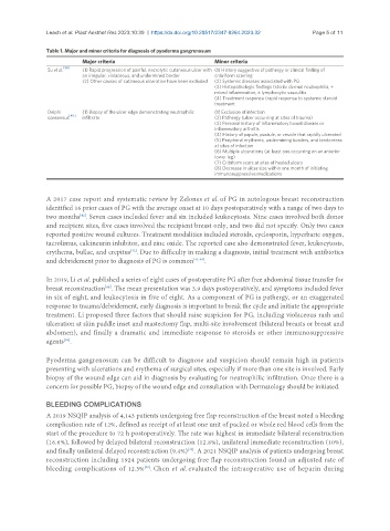
Table 1. Major and minor criteria for diagnosis of pyoderma gangrenosum
Major criteria Minor criteria
Su et al. [39] (1) Rapid progression of painful, necrolytic cutaneous ulcer with (1) History suggestive of pathergy or clinical finding of
an irregular, violaceous, and undermined border cribriform scarring
(2) Other causes of cutaneous ulceration have been excluded (2) Systemic diseases associated with PG
(3) Histopathologic findings (sterile dermal neutrophilia, ±
mixed inflammation, ± lymphocytic vasculitis
(4) Treatment response (rapid response to systemic steroid
treatment
Delphi (1) Biopsy of the ulcer edge demonstrating neutrophilic (1) Exclusion of infection
[40]
consensus infiltrate (2) Pathergy (ulcer occurring at sites of trauma)
(3) Personal history of inflammatory bowel disease or
inflammatory arthritis
(4) History of papule, pustule, or vesicle that rapidly ulcerated
(5) Peripheral erythema, undermining borders, and tenderness
at sites of infection
(6) Multiple ulcerations (at least one occurring on an anterior
lower leg)
(7) Cribiform scars at sites of healed ulcers
(8) Decrease in ulcer size within one month of initiating
immunosuppressive medications
A 2017 case report and systematic review by Zelones et al. of PG in autologous breast reconstruction
identified 16 prior cases of PG with the average onset at 10 days postoperatively with a range of two days to
two months . Seven cases included fever and six included leukocytosis. Nine cases involved both donor
[42]
and recipient sites, five cases involved the recipient breast only, and two did not specify. Only two cases
reported positive wound cultures. Treatment modalities included steroids, cyclosporin, hyperbaric oxygen,
tacrolimus, calcineurin inhibitor, and zinc oxide. The reported case also demonstrated fever, leukocytosis,
erythema, bullae, and crepitus . Due to difficulty in making a diagnosis, initial treatment with antibiotics
[42]
and debridement prior to diagnosis of PG is common [42-48] .
In 2019, Li et al. published a series of eight cases of postoperative PG after free abdominal tissue transfer for
breast reconstruction . The mean presentation was 3.9 days postoperatively, and symptoms included fever
[48]
in six of eight, and leukocytosis in five of eight. As a component of PG is pathergy, or an exaggerated
response to trauma/debridement, early diagnosis is important to break the cycle and initiate the appropriate
treatment. Li proposed three factors that should raise suspicion for PG, including violaceous rash and
ulceration at skin paddle inset and mastectomy flap, multi-site involvement (bilateral breasts or breast and
abdomen), and finally a dramatic and immediate response to steroids or other immunosuppressive
agents .
[48]
Pyoderma gangrenosum can be difficult to diagnose and suspicion should remain high in patients
presenting with ulcerations and erythema of surgical sites, especially if more than one site is involved. Early
biopsy of the wound edge can aid in diagnosis by evaluating for neutrophilic infiltration. Once there is a
concern for possible PG, biopsy of the wound edge and consultation with Dermatology should be initiated.
BLEEDING COMPLICATIONS
A 2019 NSQIP analysis of 4,143 patients undergoing free flap reconstruction of the breast noted a bleeding
complication rate of 12%, defined as receipt of at least one unit of packed or whole red blood cells from the
start of the procedure to 72 h postoperatively. The rate was highest in immediate bilateral reconstruction
(16.6%), followed by delayed bilateral reconstruction (12.8%), unilateral immediate reconstruction (10%),
and finally unilateral delayed reconstruction (9.4%) . A 2021 NSQIP analysis of patients undergoing breast
[49]
reconstruction including 1924 patients undergoing free flap reconstruction found an adjusted rate of
bleeding complications of 12.3% . Chen et al. evaluated the intraoperative use of heparin during
[30]