
Page 60 - Read Online
P. 60
Botvinkin et al. One Health Implement Res 2023;3:125-34 https://dx.doi.org/10.20517/ohir.2023.19 Page 127
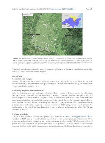
Figure 1. Spatial distribution of rabies in the Amur Region of Russia during 2018-2022. Numbers indicate animal rabies cases for
specific periods: (1) November 2018-June 2019; (2) July 2019-June 2020; (3) July 2020-June 2021; (4) July 2021-June 2022. The
asterisk indicates the first reported rabies case; the triangle indicates the human rabies case. Map insert: blue: Amur River basin; yellow:
Amur Region of Russia (ARR); black lines: borders between countries; red square: the area represented on the map.
This study aimed to detect possible ways of incursion and features of the spatial spread of rabies in ARR,
which was considered rabies-free for 45 years.
METHODS
Epidemiological analysis
Animal rabies records for 2018-2022 collected by the state epidemiological surveillance for zoonotic
infections in the ARR were used for retrospective analysis. Only animals with laboratory-confirmed rabies
were included in the study (n = 49).
Laboratory diagnosis and confirmation
After the first rabies case was reported, an active surveillance program to detect new cases was established.
During 2018-2022, the ARR Regional Veterinary Laboratory examined 1,450 brain samples of wild and
rabies-suspected domestic animals. Designated teams of hunters shot and trapped wild canids and other
game animals in different areas of ARR. Most collected wild animals did not show clinical signs of rabies or
other diseases. The direct fluorescent antibody test with FITC-conjugated anti-rabies IgG was used as the
[22]
primary method of routine diagnosis. Samples positive for RABV antigens were randomly sent for
confirmation to the Federal Reference Diagnostic Centers in Vladimir and Omsk cities, where RT-PCR was
additionally used .
[22]
Phylogenetic study
The list of RABV isolates analyzed phylogenetically is presented in Table 1 and Supplementary Table 2.
Isolation of RNA from a 10% animal brain suspension, reverse transcription, amplification of cDNA
fragments, and nucleotide sequencing were performed as described previously . Phylogenetic analysis of
[20]
partial RABV nucleoprotein (N) gene sequences (1,110 nt, positions 170-1,279 relative to Pasteur Virus
genome, GenBank no M 13215) was performed using the maximum likelihood algorithm implemented in