
Page 42 - Read Online
P. 42
Crozier-Shaw et al. Neuroimmunol Neuroinflammation 2020;7:335-44 I http://dx.doi.org/10.20517/2347-8659.2019.005 Page 3337
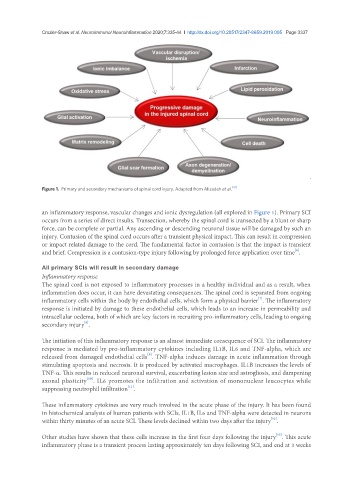
Figure 1. Primary and secondary mechanisms of spinal cord injury. Adapted from Alizadeh et al. [41]
an inflammatory response, vascular changes and ionic dysregulation (all explored in Figure 1). Primary SCI
occurs from a series of direct insults. Transection, whereby the spinal cord is transected by a blunt or sharp
force, can be complete or partial. Any ascending or descending neuronal tissue will be damaged by such an
injury. Contusion of the spinal cord occurs after a transient physical impact. This can result in compression
or impact related damage to the cord. The fundamental factor in contusion is that the impact is transient
[6]
and brief. Compression is a contusion-type injury following by prolonged force application over time .
All primary SCIs will result in secondary damage
Inflammatory response
The spinal cord is not exposed to inflammatory processes in a healthy individual and as a result, when
inflammation does occur, it can have devastating consequences. The spinal cord is separated from ongoing
[7]
inflammatory cells within the body by endothelial cells, which form a physical barrier . The inflammatory
response is initiated by damage to these endothelial cells, which leads to an increase in permeability and
intracellular oedema, both of which are key factors in recruiting pro-inflammatory cells, leading to ongoing
[8]
secondary injury .
The initiation of this inflammatory response is an almost immediate consequence of SCI. The inflammatory
response is mediated by pro-inflammatory cytokines including IL1B, IL6 and TNF-alpha, which are
[9]
released from damaged endothelial cells . TNF-alpha induces damage in acute inflammation through
stimulating apoptosis and necrosis. It is produced by activated macrophages. IL1B increases the levels of
TNF‐α. This results in reduced neuronal survival, exacerbating lesion size and astrogliosis, and dampening
[10]
axonal plasticity . IL6 promotes the infiltration and activation of mononuclear leucocytes while
[11]
suppressing neutrophil infiltration .
These inflammatory cytokines are very much involved in the acute phase of the injury. It has been found
in histochemical analysis of human patients with SCIs, IL1B, IL6 and TNF-alpha were detected in neurons
[12]
within thirty minutes of an acute SCI. These levels declined within two days after the injury .
[13]
Other studies have shown that these cells increase in the first four days following the injury . This acute
inflammatory phase is a transient process lasting approximately ten days following SCI, and end at 3 weeks