
Page 44 - Read Online
P. 44
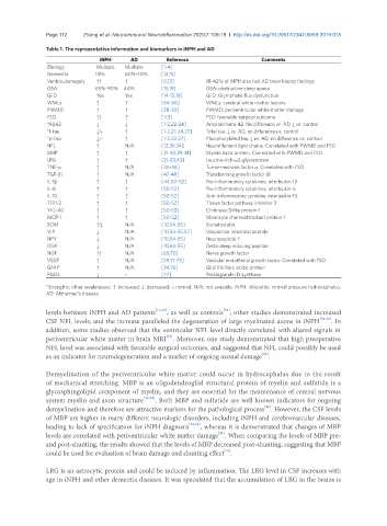
Page 112            Zhang et al. Neuroimmunol Neuroinflammation 2020;7:109-19  I  http://dx.doi.org/10.20517/2347-8659.2019.018

               Table 1. The representative information and biomarkers in iNPH and AD
                                iNPH      AD         Referencs                    Comments
                Etiology       Multiple    Multiple    [1-4]
                Dementia      10%       60%-70%    [12,15]
                Ventriculomegaly    ↑↑    ↑        [6,13]        18-42% of iNPH also had AD brain biopsy findings
                OSA           65%-90%  44%         [15,19]       OSA: obstructive sleep apnea
                GFD            Yes      Yes        [14-15,18]    GFD: Glymphatic flux dysfunction
                WMLs           ↑        ↑          [64-66]       WMLs: cerebral white matter lesions
                PWMD           ↑        ↑          [28-30]       PWMD: periventricular white matter damage
                FSO            ↑↑       ↑          [1-3]         FSO: favorable surgical outcome
                *Ab42          ↓        ↓          [1-2,22-24]    Amyloid-beta-42. No difference vs. AD, ↓ vs. control
                *t-tau         ↓/-      ↑          [1-2,21-24,27]    Total tau. ↓ vs. AD, no difference vs. control
                *p-tau         ↓/-      ↑          [1-2,22,27]    Phosphorylated tau. ↓ vs. AD, no difference vs. control
                NFL            ↑        N/A        [12,31-34]    Neurofilament light chains. Correlated with PWMD and FSO
                MBP            ↑        ↑          [31-33,39-41]    Myelin basic protein. Correlated with PWMD and FSO
                LRG            ↑        ↑          [31-33,43]    Leucine-rich-α2-glycoprotein
                TNF-α          ↑        N/A        [45-46]       Tumor-necrosis factor α. Correlated with FSO
                TGF-b1         ↑        N/A        [47-49]       Transforming growth factor β1
                IL-1b          ↑        ↑          [44,50-52]     Pro-inflammatory cytokines, interleukin-1β
                IL-6           ↑        ↑          [50-52]       Pro-inflammatory cytokines, interleukin-6
                IL-10          ↑        ↑          [50-52]       Anti-inflammatory cytokine, interleukin-10
                TFPI-2         ↑        ↑          [50-52]       Tissue factor pathway inhibitor 2
                YKL-40         ↑        ↑          [50-53]       Chitinase-3-like protein-1
                MCP-1          ↑        ↑          [50-52]       Monocyte chemoattractant protein-1
                SOM            ↑/↓      N/A        [10,54-55]    Somatostatin
                VIP            ↓        N/A        [10,54-55,57]    Vasoactive intestinal peptide
                NPY            ↓        N/A        [10,54-55]    Neuropeptide Y
                DSIP           ↓        N/A        [10,54-55]    Delta-sleep inducing peptide
                NGF            ↑↑       N/A        [69,70]       Nerve growth factor
                VEGF           ↑        N/A        [59,71-73]    Vascular endothelial growth factor. Correlated with FSO
                GFAP           ↑        N/A        [34,76]       Glial fibrillary acidic protein
                PGDS           ↓        -          [77]          Prostaglandin D synthase
               *Strengths; other weaknesses. ↑: increased; ↓: decreased; -: normal; N/A: not avaiable; iNPH: Idiopathic normal pressure hydrocephalus;
               AD: Alzheimer’s disease

                                                                      [35]
               levels between iNPH and AD patients [11,32] , as well as controls , other studies demonstrated increased
               CSF NFL levels, and the increase paralleled the degeneration of large myelinated axons in iNPH [31,36] . In
               addition, some studies observed that the ventricular NFL level directly correlated with altered signals in
                                                    [37]
               periventricular white matter in brain MRI . Moreover, one study demonstrated that high preoperative
               NFL level was associated with favorable surgical outcomes, and suggested that NFL could possibly be used
                                                                                  [38]
               as an indicator for neurodegeneration and a marker of ongoing axonal damage .

               Demyelination of the periventricular white matter could occur in hydrocephalus due to the result
               of mechanical stretching. MBP is an oligodendroglial structural protein of myelin and sulfatide is a
               glycosphingolipid component of myelin, and they are essential for the maintenance of central nervous
               system myelin and axon structure [32,39] . Both MBP and sulfatide are well known indicators for ongoing
                                                                                  [40]
               demyelination and therefore are attractive markers for the pathological process . However, the CSF levels
               of MBP are higher in many different neurologic disorders, including iNPH and cerebrovascular diseases,
               leading to lack of specification for iNPH diagnosis [32,36] , whereas it is demonstrated that changes of MBP
                                                                    [41]
               levels are correlated with periventricular white matter damage . When comparing the levels of MBP pre-
               and post-shunting, the results showed that the levels of MBP decreased post-shunting, suggesting that MBP
               could be used for evaluation of brain damage and shunting effect .
                                                                      [42]
               LRG is an astrocytic protein and could be induced by inflammation. The LRG level in CSF increases with
               age in iNPH and other dementia diseases. It was speculated that the accumulation of LRG in the brains is