
Page 88 - Read Online
P. 88
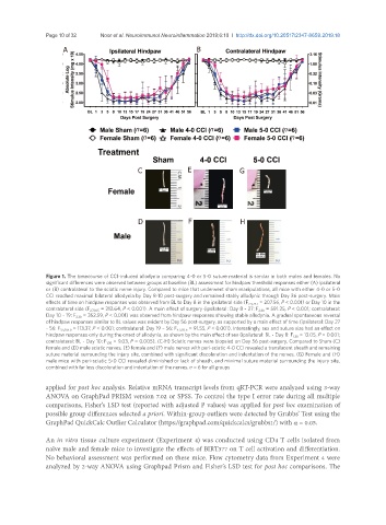
Page 10 of 32                 Noor et al. Neuroimmunol Neuroinflammation 2019;6:10  I  http://dx.doi.org/10.20517/2347-8659.2019.18

                     A                                        B


                                           n                      n                     n
                                             n                      n                      n


                                            C              E               G


                                            D              F               H


               Figure 1. The timecourse of CCI-induced allodynia comparing 4-0 or 5-0 suture material is similar in both males and females. No
               significant differences were observed between groups at baseline (BL) assessment for hindpaw threshold responses either (A) ipsilateral
               or (B) contralateral to the sciatic nerve injury. Compared to mice that underwent sham manipulations, all mice with either 4-0 or 5-0
               CCI reached maximal bilateral allodynia by Day 8-10 post-surgery and remained stably allodynic through Day 36 post-surgery. Main
               effects of time on hindpaw responses was observed from BL to Day 8 in the ipsilateral side (F 2.4,74.7  = 207.56, P < 0.001) or Day 10 in the
               contralateral side (F 2.7,81.5  = 212.64, P < 0.001). A main effect of surgery (ipsilateral: Day 8 - 27: F 2,30  = 591.25, P < 0.001; contralateral:
               Day 10 - 19: F 2,30  = 352.59, P < 0.001) was observed from hindpaw responses showing stable allodynia. A gradual spontaneous reversal
               of hindpaw responses similar to BL values was evident by Day 56 post-surgery, as supported by a main effect of time (ipsilateral: Day 27
               - 56: F 3.5,105.0  = 113.37, P < 0.001; contralateral: Day 19 – 56: F 4.3,131.6  = 91.55, P < 0.001). Interestingly, sex and suture size had an effect on
               hindpaw responses only during the onset of allodynia, as shown by the main effect of sex (ipsilateral: BL - Day 8: F 1,30  = 13.05, P = 0.001;
               contralateral: BL - Day 10: F 1,30  = 9.03, P = 0.005). (C-H) Sciatic nerves were biopsied on Day 56 post-surgery. Compared to Sham (C)
               female and (D) male sciatic nerves, (E) female and (F) male nerves with peri-sciatic 4-0 CCI revealed a translucent sheath and remaining
               suture material surrounding the injury site, combined with significant discoloration and indentation of the nerves. (G) Female and (H)
               male mice with peri-sciatic 5-0 CCI revealed diminished or lack of sheath, and minimal suture material surrounding the injury site,
               combined with far less discoloration and indentation of the nerves. n = 6 for all groups

               applied for post hoc analysis. Relative mRNA transcript levels from qRT-PCR were analyzed using 3-way
               ANOVA on GraphPad PRISM version 7.02 or SPSS. To control the type I error rate during all multiple
               comparisons, Fisher’s LSD test (reported with adjusted P values) was applied for post hoc examination of
               possible group differences selected a priori. Within-group outliers were detected by Grubbs’ Test using the
               GraphPad QuickCalc Outlier Calculator (https://graphpad.com/quickcalcs/grubbs1/) with α = 0.05.


               An in vitro tissue culture experiment (Experiment 4) was conducted using CD4 T cells isolated from
               naïve male and female mice to investigate the effects of BIRT377 on T cell activation and differentiation.
               No behavioral assessment was performed on these mice. Flow cytometry data from Experiment 4 were
               analyzed by 2-way ANOVA using Graphpad Prism and Fisher’s LSD test for post hoc comparisons. The