
Page 31 - Read Online
P. 31
Kaya et al. Neuroimmunol Neuroinflammation 2019;6:5 I http://dx.doi.org/10.20517/2347-8659.2018.70 Page 5 of 15
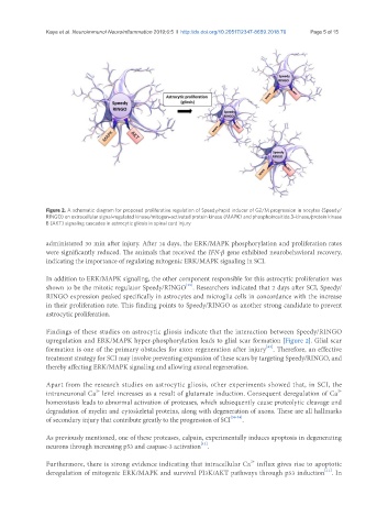
Figure 2. A schematic diagram for proposed proliferative regulation of Speedy/rapid inducer of G2/M progression in oocytes (Speedy/
RINGO) on extracellular signal-regulated kinase/mitogen-activated protein kinase (MAPK) and phosphoinositide 3-kinase/protein kinase
B (AKT) signaling cascades in astrocytic gliosis in spinal cord injury
administered 30 min after injury. After 14 days, the ERK/MAPK phosphorylation and proliferation rates
were significantly reduced. The animals that received the IFN-β gene exhibited neurobehavioral recovery,
indicating the importance of regulating mitogenic ERK/MAPK signaling in SCI.
In addition to ERK/MAPK signaling, the other component responsible for this astrocytic proliferation was
[15]
shown to be the mitotic regulator Speedy/RINGO . Researchers indicated that 2 days after SCI, Speedy/
RINGO expression peaked specifically in astrocytes and microglia cells in concordance with the increase
in their proliferation rate. This finding points to Speedy/RINGO as another strong candidate to prevent
astrocytic proliferation.
Findings of these studies on astrocytic gliosis indicate that the interaction between Speedy/RINGO
upregulation and ERK/MAPK hyper-phosphorylation leads to glial scar formation [Figure 2]. Glial scar
[63]
formation is one of the primary obstacles for axon regeneration after injury . Therefore, an effective
treatment strategy for SCI may involve preventing expansion of these scars by targeting Speedy/RINGO, and
thereby affecting ERK/MAPK signaling and allowing axonal regeneration.
Apart from the research studies on astrocytic gliosis, other experiments showed that, in SCI, the
2+
2+
intraneuronal Ca level increases as a result of glutamate induction. Consequent deregulation of Ca
homeostasis leads to abnormal activation of proteases, which subsequently cause proteolytic cleavage and
degradation of myelin and cytoskeletal proteins, along with degeneration of axons. These are all hallmarks
of secondary injury that contribute greatly to the progression of SCI [30-34] .
As previously mentioned, one of these proteases, calpain, experimentally induces apoptosis in degenerating
[13]
neurons through increasing p53 and caspase-3 activation .
2+
Furthermore, there is strong evidence indicating that intracellular Ca influx gives rise to apoptotic
[11]
deregulation of mitogenic ERK/MAPK and survival PI3K/AKT pathways through p53 induction . In