
Page 335 - Read Online
P. 335
Choi et al. Neuroimmunol Neuroinflammation 2018;5:42 I http://dx.doi.org/10.20517/2347-8659.2018.47 Page 7 of 14
E
C
A
D
B
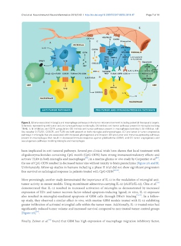
Figure 2. Glioma-associated microglia and macrophage pathways in the tumor microenvironment including potential therapeutic targets.
Schematic representing anti-tumor and pro-tumor pathways in microglia. (A) Intrinsic anti-tumor pathways present in microglia involving
TRAIL, IL-1β inhibition, and CD74 upregulation; (B) Intrinsic anti-tumor pathways present in macrophages involving IL-1β inhibition. toll-
like receptor 3 (TLR3), CX3CR1, and TLR9 are both present on both microglia and macrophages; (C) pro-tumor or tumor-progressive
pathways in microglia that are associated with increased gliomagenesis and invasion; (D) pro-tumor and immunosuppressing pathways
present in macrophages that result in decreased immune response against glioblastoma (GBM); and (E) tumor angiogenesis and
vasculogenesis pathways involving microglia and macrophages
been implicated in anti-tumoral pathways. Several pre-clinical trials have shown that local treatment with
oligodeoxynucleotides containing CpG motifs (CpG-ODN) have strong immunostimulatory effects and
[66]
[67]
activate TLR9 in both microglia and macrophages ; in a murine glioma in vivo study by Carpentier et al. ,
the use of CpG-ODN resulted in decreased tumor size without toxicity to brain parenchyma [Figure 2A and B].
Unfortunately, follow-up studies in humans including a phase II trial did not show significant progression
free survival or radiological response in patients treated with CpG-ODN [68,69] .
More promisingly, another study demonstrated the importance of IL-12 in the modulation of microglial anti-
tumor activity in mouse models. Using recombinant adenovirus-carrying IL-12 (rAAV2/IL-12), Chiu et al. [70]
demonstrated that IL-12 resulted in increased activation of microglia as demonstrated by increased
expression of ED1 and tumor necrosis factor-related apoptosis-inducing ligand; in vitro, IL-12 exposure
also resulted in microglial-mediated apoptosis of GBM cells through DR4/5 binding [70,71] . In a follow-
up study, they observed a similar effect in vivo, with murine GBM models treated with IL-12 exhibiting
greater infiltration of activated microglial cells within the tumor mass. Additionally, IL-12 treated mice had
significantly reduced tumor volume and increased survival compared to non-treated tumor control groups
[70]
[Figure 2A] .
Finally, Zeiner et al. found that GBM has high expression of macrophage migration inhibitory factor,
[72]