
Page 202 - Read Online
P. 202
Rana et al. Takayasu’s arteritis
A B
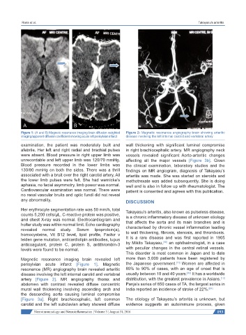
Figure 1: (A and B) Magnetic resonance imaging brain diffusion weighted Figure 2: Magnetic resonance angiography brain showing arteritic
imaging/apparent diffusion coefficient showing acute left perisylvian infarct disease involving the left internal carotid and vertebral artery
examination, the patient was moderately built and wall thickening with significant luminal compromise
afebrile. Her left and right radial and brachial pulses in right brachiocephalic artery. MR angiography neck
were absent. Blood pressure in right upper limb was vessels revealed significant Aorto-arteritic changes
unrecordable and left upper limb was 120/70 mmHg. affecting all the major vessels [Figure 3b]. Given
Blood pressure recorded in the lower limbs was the clinical examination, laboratory studies and the
130/80 mmHg on both the sides. There was a thrill findings on MR angiogram, diagnosis of Takayasu’s
associated with a bruit over the right carotid artery. All arteritis was made. She was started on steroids and
the lower limb pulses were felt. She had wernicke’s methotrexate was added subsequently. She is doing
aphasia, no facial asymmetry, limb power was normal. well and is also in follow up with rheumatologist. The
Cardiovascular examination was normal. There were patient is consented and agrees with this publication.
no renal vascular bruits and optic fundi did not reveal
any abnormality. DISCUSSION
Her erythrocyte segmentation rate was 50 mm/h, total Takayasu’s arteritis, also known as pulseless disease,
counts 5,200 cells/µL, C-reactive protein was positive, is a chronic inflammatory disease of unknown etiology
and chest X-ray was normal. Electrocardiogram and that affects the aorta and its main branches and is
holter study was within normal limit. Echo cardiography
revealed normal study. Serum lipoprotein(a), characterised by chronic vessel inflammation leading
homocysteine, Vit B12 level, lipid profile, Factor v to wall thickening, fibrosis, stenosis, and thrombosis.
leiden gene mutation, anticardiolipin antibodies, lupus It is a rare disease and was first reported in 1905
[10]
anticoagulant, protein C, protein S, antithrombin-3 by Mikito Takayasu, an ophthalmologist, in a case
levels were found to be normal. with peculiar changes in the central retinal vessels.
This disorder is most common in Japan and to date
Magnetic resonance imaging brain revealed left more than 5,000 patients have been registered by
[11]
perisylvian acute infarct [Figure 1]. Magnetic the Japanese government. Women are affected in
resonance (MR) angiography brain revealed arteritic 80% to 90% of cases, with an age of onset that is
[12]
disease involving the left internal carotid and vertebral usually between 10 and 40 years. It has a worldwide
artery [Figure 2]. MR angiography thorax and distribution, with the greatest prevalence in Asians.
[13]
abdomen with contrast revealed diffuse concentric Panja’s series of 650 cases of TA, the largest series in
mural wall thickening involving ascending arch and India reported an incidence of stroke of 22%. [14]
the descending aorta causing luminal compromise
[Figure 3a]. Right brachiocephalic, left common The etiology of Takayasu’s arteritis is unknown, but
carotid and the left subclavian artery showed diffuse evidence suggests an autoimmune process, given
Neuroimmunology and Neuroinflammation ¦ Volume 3 ¦ August 31, 2016 193