
Page 390 - Read Online
P. 390
Page 6 of 10 Gharagozloo et al. Mini-invasive Surg 2021;5:39 https://dx.doi.org/10.20517/2574-1225.2021.74
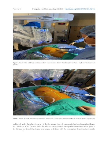
Figure 6. Patient in the left lateral decubitus position. Trocar sites are shown. The robot (da Vinci Xi) is brought over the head of the
patient.
Figure 7. Patient in the left lateral decubitus position. The retractor (arrow) which is fixed to the bed is used to retract the lung inferiorly.
and the rib under the subclavian artery is divided using a 6-mm thoracoscopic Kerrison bone cutter (Depuy
Inc., Raynham, MA). The area under the subclavian artery, which corresponds with the subclavian grove, is
the thinnest portion of the rib and is amenable to division with the bone cutter. The rib’s division at its