
Page 138 - Read Online
P. 138
Ackerman et al. Mini-invasive Surg 2021;5:14 https://dx.doi.org/10.20517/2574-1225.2021.02 Page 9 of 19
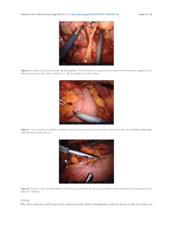
Figure 6. Greater curve dissection with individual ligation of the short gastric vessels is performed with the retrogastric approach. SG:
Short gastric vessels; RC: right crural pillar; LC: left crural pillar; SA: splenic artery.
Figure 7. The stomach is oriented for conduit creation by retracting the tip of the fundus towards the apex of the left hemidiaphragm
with the robotic assist (arm 4).
Figure 8. The first robotic vascular stapler is fired 5-6 cm proximal to the pylorus to include the lesser omentum and ending just on the
stomach. P: Pylorus.
Closing
The liver retractor and its port are removed under direct visualization and the fascia of the port sites are