
Page 862 - Read Online
P. 862
Page 2 of 7 Herbella et al. Mini-invasive Surg 2020;4:82 I http://dx.doi.org/10.20517/2574-1225.2020.84
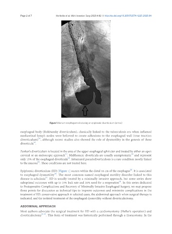
Figure 1. Barium esophagram disclosing an epiphrenic diverticulum (arrow)
esophageal body (Rokitansky diverticulum), classically linked to the tuberculosis era when inflamed
mediastinal lymph nodes were believed to create adhesions to the esophageal wall (true traction
[2]
diverticulum) , although recent studies also showed the role of dysmotility in the genesis of these
[3]
diverticula .
Zenker’s diverticulum is located in the area of the upper esophageal sphincter and treated by either an open
[1]
cervical or an endoscopic approach . Midthoracic diverticula are usually asymptomatic and represent
[3]
[4]
only 15% of the esophageal diverticula . Intramural pseudodiverticulosis is a rare condition mostly linked
[5]
to the mucosa . These conditions are not treated here.
[2]
Epiphrenic diverticulum (ED) [Figure 1] occurs within the distal 10 cm of the esophagus . It is associated
[6]
to esophageal dysmotility . The most common named esophageal motility disorder linked to this
disease is achalasia . ED is usually treated by a minimally invasive approach, but some series show
[7]
[8]
suboptimal outcomes with up to 23% leak rate and 20% need for a reoperation . In this series dedicated
to Postoperative Complications and Recovery of Minimally Invasive Esophageal Surgery, we may propose
three points for discussion as technical tips to improve outcomes and minimize complications in the
treatment of ED: conservative approach in selected cases, the abdominal approach when surgical therapy is
indicated, and the isolated treatment of the esophageal dysmotility without diverticulectomy.
ABDOMINAL APPROACH
Most authors advocate the surgical treatment for ED with a cardiomyotomy (Heller’s operation) and
diverticulectomy [8-10] . This form of treatment was historically performed through a thoracotomy. In the