
Page 556 - Read Online
P. 556
Page 4 of 12 Belluschi et al. Mini-invasive Surg 2020;4:58 I http://dx.doi.org/10.20517/2574-1225.2020.48
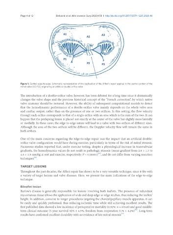
Figure 1. Central edge-to-edge. Schematic representation of the application of the Alfieri’s repair applied in the central portion of the
mitral valve (A2-P2), originating an artificial double-orifice valve
The introduction of a double-orifice valve, however, has been debated for a long time since it dramatically
changes the valve shape and the previous historical concept of the “French correction”, by which native
valve anatomy should be restored. However, the ability of subsequent computational models to detect
that the hemodynamic performance of a double-orifice valve mainly depends on the whole valve area
and cardiac output, rather than on the presence of one or two orifices. In this setting, the flow velocity
through each orifice corresponds to that of a single orifice with an area which is the sum of the two. It can
happen that the prolapsing lesion is placed not exactly at the center of the valve but slightly more laterally
or medially. In these cases, the edge-to-edge suture will lead to a valve with two orifices of different sizes.
Although the area of the two orifices will be different, the Doppler velocity flow will remain the same in
both orifices.
One of the main concerns regarding the edge-to-edge repair was the impact that an artificial double-
orifice valve configuration would have during exercise, particularly in terms of the risk of mitral stenosis.
Numerous studies reported that, under exercise testing, despite a physiological increase in transvalvular
gradients, the hemodynamics values do not result in pathologic stenosis (mean gradient from 2.8 ± 1.3 to
[21]
4.6 ± 1.9 mmHg at rest and exercise, respectively; P < 0.00001) , and do not differ from varying resection
[22]
techniques .
TARGET LESIONS
Throughout the past decades, the Alfieri repair has shown to be a very versatile technique, since it fits with
a variety of target lesions and valve diseases. Here, we present the main indications of the edge-to-edge
technique.
Bileaflet lesion
Barlow’s disease is generally responsible for lesions involving both leaflets. The presence of redundant
myxomatous tissue allows the application of wide and deep edge-to-edge stitches, thus reducing the leaflets’
height. In addition, converse to longer procedures targeting the chordal/papillary muscle apparatus, it can
be easily and quickly performed, thus reducing ischemic time while still achieving excellent results. The
first published data showed a low incidence of perioperative mortality (0.92%; n = 6/648) and good middle-
[23]
term clinical outcome (5-year survival 92% ± 4.5%; freedom from reoperation 91% ± 4.2%) . Long-term
[24]
results have confirmed excellent durability with no evidence of late mitral stenosis .