
Page 248 - Read Online
P. 248
Fontan et al. Mini-invasive Surg 2020;4:29 I http://dx.doi.org/10.20517/2574-1225.2020.09 Page 3 of 11
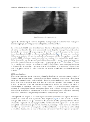
Figure 1. Esophagram showing a hiatal hemia
augment this anatomic region. Moreover, the phreno-esophageal ligament anchors the distal esophagus to
[13]
the crural diaphragm, preventing excessive sliding during respiratory cycles .
The development of GERD is usually multifactorial. A failure of the anti-reflux barrier that comprises the
LES and the crural muscles of the hiatus are common factors in the pathophysiology. Curiously, in a cohort
[14]
that included 1659 patients with foregut symptoms, Ayazi et al. was able to demonstrate that the presence
of a mechanically defective LES, as well as concomitant hiatal hernias [Figure 1], became more prevalent as
BMI increased. Indirectly, LES function can be affected by extrinsic variables. Obese patients’ susceptibility
to develop GERD is intimately related to these indirect variables, which include higher gastric capacity
(higher distensibility and disruption of muscle fibers), increased intra-gastric pressure, and augmented
[18]
positive intra-abdominal pressure as well as negative intra-thoracic pressure [15-17] . Herbella et al. found
that for each five-point increment in an obese patient’s BMI, the DeMeester score was expected to increase
by three units. Furthermore, from a hormonal standpoint, irregularities in the secretion of adiponectin and
leptin from adipose tissue cells has been proposed as a potential nexus between obesity and esophageal
metaplasia [4,19] .
GERD complications
GERD complications are related to excessive reflux of acid and pepsin, which can result in necrosis of
the mucosa. The amount of injury occasionally outweighs the remodeling capacity of the cellular lining,
leading to erosions and ulcers, a condition which is defined as erosive esophagitis. A potential complication
seen in GERD patients with esophagitis is the development of peptic strictures. These strictures can occur
secondary to persistent injury. Scar tissue forms due to chronic necrosis and inflammation, leading to
variable degrees of physiologic contraction of collagen fibers. This phenomenon can cause significant
narrowing of the esophageal lumen at the esophago-gastric outlet. This type of benign stricture is usually
short segment, circumferential, and amenable to therapeutic dilations for patency restoration. Fortunately,
[20]
the incidence of strictures has significantly declined since the beginning of the PPI era .
Certain patients can progress to develop metaplastic columnar epithelium which replaces the stratified
squamous epithelium that normally lines the distal esophagus [Figure 2]. This is defined as Barrett’s
esophagus (BE), and the endoscopic prevalence of this phenomenon in the general population is between
[21]
0.5% and 2%. For patients with underlying GERD, the prevalence rises to as high as 15% . In fact, erosive
esophagitis is considered an independent risk factor for BE, conferring a fivefold increased risk in a five-
[22]
year follow-up period . Not surprisingly, the prevalence of BE in the obese population can be as high
as 40%. These alarming rates are some of the reasons why current trends favor aggressive preoperative