
Page 13 - Read Online
P. 13
Cortes-Cerisuelo et al. Mini-invasive Surg 2019;3:1 I http://dx.doi.org/10.20517/2574-1225.2018.60 Page 5 of 10
A B
C D
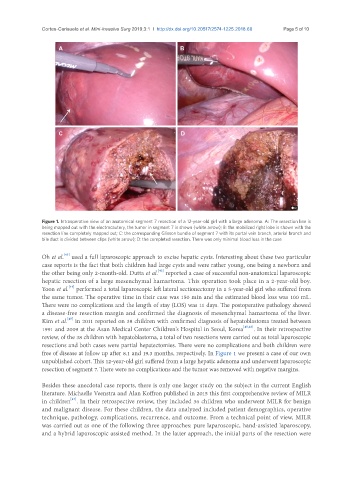
Figure 1. Intraoperative view of an anatomical segment 7 resection of a 12-year-old girl with a large adenoma. A: The resection line is
being mapped out with the electrocautery, the tumor in segment 7 is shown (white arrow); B: the mobilized right lobe is shown with the
resection line completely mapped out; C: the corresponding Glisson bundle of segment 7 with its portal vein branch, arterial branch and
bile duct is divided between clips (white arrow); D: the completed resection. There was only minimal blood loss in the case
[43]
Oh et al. used a full laparoscopic approach to excise hepatic cysts. Interesting about these two particular
case reports is the fact that both children had large cysts and were rather young, one being a newborn and
[42]
the other being only 2-month-old. Dutta et al. reported a case of successful non-anatomical laparoscopic
hepatic resection of a large mesenchymal hamartoma. This operation took place in a 2-year-old boy.
[44]
Yoon et al. performed a total laparoscopic left lateral sectionectomy in a 5-year-old girl who suffered from
the same tumor. The operative time in their case was 150 min and the estimated blood loss was 100 mL.
There were no complications and the length of stay (LOS) was 11 days. The postoperative pathology showed
a disease-free resection margin and confirmed the diagnosis of mesenchymal hamartoma of the liver.
[45]
Kim et al. in 2011 reported on 38 children with confirmed diagnosis of hepatoblastoma treated between
1991 and 2009 at the Asan Medical Center Children’s Hospital in Seoul, Korea [45,46] . In their retrospective
review, of the 38 children with hepatoblastoma, a total of two resections were carried out as total laparoscopic
resections and both cases were partial hepatectomies. There were no complications and both children were
free of disease at follow up after 8.1 and 19.3 months, respectively. In Figure 1 we present a case of our own
unpublished cohort. This 12-year-old girl suffered from a large hepatic adenoma and underwent laparoscopic
resection of segment 7. There were no complications and the tumor was removed with negative margins.
Besides these anecdotal case reports, there is only one larger study on the subject in the current English
literature. Michaelle Veenstra and Alan Koffron published in 2015 this first comprehensive review of MILR
[47]
in children . In their retrospective review, they included 36 children who underwent MILR for benign
and malignant disease. For these children, the data analyzed included patient demographics, operative
technique, pathology, complications, recurrence, and outcome. From a technical point of view, MILR
was carried out as one of the following three approaches: pure laparoscopic, hand-assisted laparoscopy,
and a hybrid laparoscopic assisted method. In the latter approach, the initial parts of the resection were