
Page 91 - Read Online
P. 91
Jani. Mini-invasive Surg 2018;2:14 I http://dx.doi.org/10.20517/2574-1225.2018.08 Page 3 of 9
Figure 1. Port positions
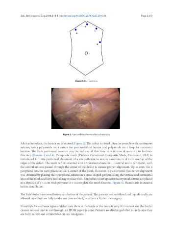
Figure 2. Para-umbilical hernia after adhesiolysis
After adhesiolysis, the hernia sac is excised [Figure 2]. The defect is closed intra-corporeally with continuous
sutures, using polyamide no 1 suture for para-umbilical hernia and polyamide no 1 loop for incisional
hernias. The intra-peritoneal pressure may be reduced at this time to 8-10 mm of mercury to facilitate
this step [Figures 3 and 4]. Composite mesh (Parietex Optimized Composite Mesh, Medtronic, USA) is
introduced for intra-peritoneal placement of a size sufficient to ensure a minimum of 5 cm overlap of the
edges of the defect. The mesh is first oriented with 5 transfascial sutures - 1 central and 4 peripheral, with
the central sutures passed through the center of the defect to ensure proper alignment. Up to 2010, the 4
peripheral sutures were placed at the 4 corner of the mesh. However, we discovered that better alignment
was obtained by placing the 4 peripheral sutures in a cross shaped pattern, along the vertical and horizontal
axes of the mesh and have been doing so since then. Thereafter, interrupted intracorporeal sutures are placed
at a distance of 1-1.5 cm with polyester 2-0 to complete the mesh fixation [Figure 5]. Hemostasis is ensured
before desufflation.
The Ryle’s tube is removed before extubation of the patient. The patients are mobilized and liquids orally are
allowed once they are fully awake and non-sedated, usually 3-4 h after the surgery.
If multiple Swiss cheese types of defects are there in the fascia or the fascia is very thinned out and the fascial
closure sutures tend to cut through, an IPOM repair is done. Patients are discharged after 24-48 h once they
are fully mobile and comfortable on oral analgesics.