
Page 56 - Read Online
P. 56
Ogura et al. Mini-invasive Surg 2018;2:8 I http://dx.doi.org/10.20517/2574-1225.2017.48 Page 3 of 4
A B
C D
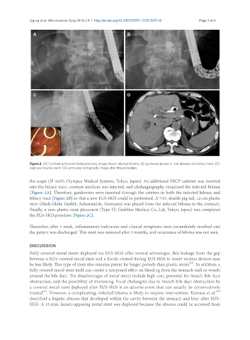
Figure 2. (A) Contrast-enhanced cholangioscopy image shows infected biloma; (B) guidewire placed in liver abscess and biliary tract; (C)
deployed double stent; (D) computed tomography image after this procedure
the scope (JF 260V; Olympus Medical Systems, Tokyo, Japan). An additional ERCP catheter was inserted
into the biliary tract, contrast medium was injected, and cholangiography visualized the infected biloma
[Figure 2A]. Therefore, guidewires were inserted through the cateters in both the infected biloma and
biliary tract [Figure 2B] so that a new EUS-HGS could be performed. A 7-Fr, double pig tail, 12-cm plastic
stent (Medi-Globe GmbH; Achenmühle, Germany) was placed from the infected biloma to the stomach.
Finally, a new, plastic stent placement (Type IT; Gadelius Medical Co, Ltd, Tokyo, Japan) was completed
the EUS-HGS proedure [Figure 2C].
Thereafter, after 1 week, inflammatory indicators and clinical symptoms were immediately resolved and
the patient was discharged. This stent was removed after 2 months, and recurrence of biloma was not seen.
DISCUSSION
Fully covered metal stents deployed via EUS-HGS offer several advantages. Bile leakage from the gap
between a fully covered metal stent and a fistula created during EUS-HGS to insert various devices may
[11]
be less likely. This type of stent also remains patent for longer periods than plastic stents . In addition, a
fully covered metal stent itself can confer a tamponed effect on bleeding from the stomach wall or vessels
around the bile duct. The disadvantages of metal stents include high cost, potential for branch bile duct
obstruction, and the possibility of shortening. Focal cholangitis due to branch bile duct obstruction by
a covered metal stent deployed after EUS-HGS is an adverse event that can usually be conservatively
[12]
[12]
treated . However, a complicating, infected biloma is likely to require intervention. Kumata et al.
described a hepatic abscess that developed within the cavity between the stomach and liver after EUS-
HGS. A 15-mm, lumen-apposing metal stent was deployed because the abscess could be accessed from