
Page 367 - Read Online
P. 367
Page 4 of 12 Chen et al. Mini-invasive Surg 2018;2:43 I http://dx.doi.org/10.20517/2574-1225.2018.42
A B
C D
E F
G
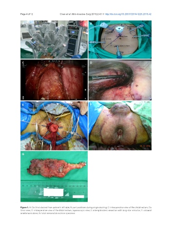
Figure 1. A: Da Vinci docked from patient’s left side; B: port positions during single docking; C: intraoperative view of the distal rectum, Da
Vinci view; D: intraoperative view of the distal rectum, laparoscopic view; E: intersphincteric resection with long-star retractor; F: coloanal
anastomosis done; G: total mesorectal excision specimen