
Page 60 - Read Online
P. 60
Shichijo et al. Mini-invasive Surg 2022;6:19 https://dx.doi.org/10.20517/2574-1225.2021.121 Page 5 of 13
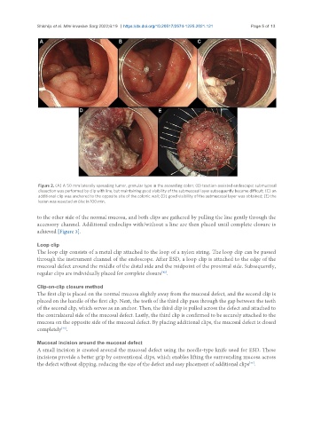
Figure 2. (A) A 50 mm laterally spreading tumor, granular type in the ascending colon; (B) traction-assisted endoscopic submucosal
dissection was performed by clip with line, but maintaining good visibility of the submucosal layer subsequently became difficult; (C) an
additional clip was anchored to the opposite site of the colonic wall; (D) good visibility of the submucosal layer was obtained; (E) the
lesion was resected en bloc in 100 min.
to the other side of the normal mucosa, and both clips are gathered by pulling the line gently through the
accessory channel. Additional endoclips with/without a line are then placed until complete closure is
achieved [Figure 3].
Loop clip
The loop clip consists of a metal clip attached to the loop of a nylon string. The loop clip can be passed
through the instrument channel of the endoscope. After ESD, a loop clip is attached to the edge of the
mucosal defect around the middle of the distal side and the midpoint of the proximal side. Subsequently,
[42]
regular clips are individually placed for complete closure .
Clip-on-clip closure method
The first clip is placed on the normal mucosa slightly away from the mucosal defect, and the second clip is
placed on the handle of the first clip. Next, the teeth of the third clip pass through the gap between the teeth
of the second clip, which serves as an anchor. Then, the third clip is pulled across the defect and attached to
the contralateral side of the mucosal defect. Lastly, the third clip is confirmed to be securely attached to the
mucosa on the opposite side of the mucosal defect. By placing additional clips, the mucosal defect is closed
completely .
[43]
Mucosal incision around the mucosal defect
A small incision is created around the mucosal defect using the needle-type knife used for ESD. These
incisions provide a better grip by conventional clips, which enables lifting the surrounding mucosa across
[44]
the defect without slipping, reducing the size of the defect and easy placement of additional clips .