
Page 35 - Read Online
P. 35
Page 4 of 7 Takamaru et al. Mini-invasive Surg 2022;6:15 https://dx.doi.org/10.20517/2574-1225.2021.120
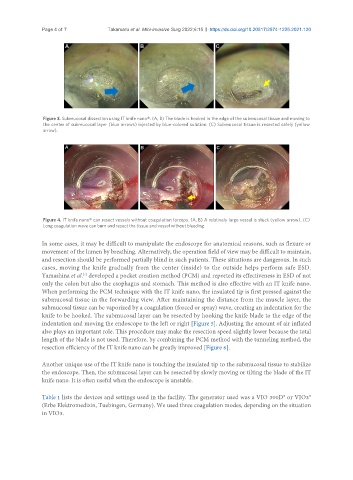
Figure 3. Submucosal dissection using IT knife nano®. (A, B) The blade is hooked in the edge of the submucosal tissue and moving to
the center of submucosal layer (blue arrows) injected by blue-colored solution. (C) Submucosal tissue is resected safely (yellow
arrow).
Figure 4. IT knife nano® can resect vessels without coagulation forceps. (A, B) A relatively large vessel is stuck (yellow arrow). (C)
Long coagulation wave can burn and resect the tissue and vessel without bleeding.
In some cases, it may be difficult to manipulate the endoscope for anatomical reasons, such as flexure or
movement of the lumen by breathing. Alternatively, the operation field of view may be difficult to maintain,
and resection should be performed partially blind in such patients. These situations are dangerous. In such
cases, moving the knife gradually from the center (inside) to the outside helps perform safe ESD.
Yamashina et al. developed a pocket creation method (PCM) and reported its effectiveness in ESD of not
[7]
only the colon but also the esophagus and stomach. This method is also effective with an IT knife nano.
When performing the PCM technique with the IT knife nano, the insulated tip is first pressed against the
submucosal tissue in the forwarding view. After maintaining the distance from the muscle layer, the
submucosal tissue can be vaporized by a coagulation (forced or spray) wave, creating an indentation for the
knife to be hooked. The submucosal layer can be resected by hooking the knife blade to the edge of the
indentation and moving the endoscope to the left or right [Figure 5]. Adjusting the amount of air inflated
also plays an important role. This procedure may make the resection speed slightly lower because the total
length of the blade is not used. Therefore, by combining the PCM method with the tunneling method, the
resection efficiency of the IT knife nano can be greatly improved [Figure 6].
Another unique use of the IT knife nano is touching the insulated tip to the submucosal tissue to stabilize
the endoscope. Then, the submucosal layer can be resected by slowly moving or tilting the blade of the IT
knife nano. It is often useful when the endoscope is unstable.
Table 1 lists the devices and settings used in the facility. The generator used was a VIO 300D® or VIO3®
(Erbe Elektromedizin, Tuebingen, Germany). We used three coagulation modes, depending on the situation
in VIO3.