
Page 27 - Read Online
P. 27
Page 4 of 8 Kato et al. Mini-invasive Surg 2022;6:10 https://dx.doi.org/10.20517/2574-1225.2021.124
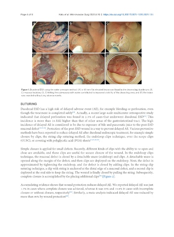
Figure 1. Duodenal ESD using the water pressure method: (A) a 40 mm flat elevated lesion was found in the descending duodenum; (B,
C) mucosal incision; (D, E) hitting the submucosa with water contributed to improved visibility of the dissecting area; and (F) the lesion
was resected without any adverse events.
SUTURING
Duodenal ESD has a high risk of delayed adverse event (AE), for example bleeding or perforation, even
though the treatment is completed safely . Actually, a recent large-scale multicenter retrospective study
[33]
indicated that delayed perforation was found in 2.3% of cases that underwent duodenal ESD . This
[23]
incidence is more than 10-fold higher than that of other areas of the gastrointestinal tract. The high
incidence of delayed AE is considered to be due to exposure of bile and pancreatic juice to the post-ESD
mucosal defect [18,33-35] . Protection of the post-ESD wound is a way to prevent delayed AE. Various preventive
methods have been reported to reduce delayed AE after duodenal endoscopic treatment, for example simple
closure by clips, the string-clip suturing method, the endoloop-clips technique, over-the-scope clips
(OTSC), or covering with polyglycolic acid (PGA) sheets [19,21,36-39] .
Simple closure is applied for small defects. Recently, different kinds of clips with the ability to re-open and
close are available, and these clips are useful for secure closure of the wound. In the endoloop-clips
technique, the mucosal defect is closed by a detachable snare (endoloop) and clips. A detachable snare is
opened along the margin of the defect, and then clips are deployed on the endoloop. Next, the defect is
approximated by tightening the endoloop, and the defect is closed by adding clips. In the string-clip
suturing technique, a clip with string is anchored at the distal edge of a mucosal defect, and a second clip is
deployed at the oral side to keep the string. The wound is finally closed by pulling the string. Subsequently,
[39]
complete closure is accomplished by the placing additional clips [Figure 2].
Accumulating evidence shows that wound protection reduces delayed AE. We reported delayed AE was just
1.7% in cases where complete closure was achieved, whereas it was 25% and 15.6% in cases with incomplete
closure or without closure, respectively . Similarly, a meta-analysis indicated delayed AE was reduced by
[36]
more than 80% by wound protection .
[40]