
Page 20 - Read Online
P. 20
Hayashi et al. Mini-invasive Surg 2022;6:7 https://dx.doi.org/10.20517/2574-1225.2021.125 Page 5 of 8
Figure 4. The advantage of a distant entrance. (A) When the initial mucosal incision as the entrance to the pocket is close to the tumor,
the mucosal edge will be rolled. (B) A distant initial incision applies more upward traction and prevents rolling. The ideal distance is
14 mm, the same as twice the diameter of the small-caliber-tip transparent hood orifice.
Figure 5. Successive short dissections. (A) Hooking the submucosa slightly in the direction of cut. (B) When pressing the pedal of the
diathermy unit, the hooked submucosa is cut. (C) Performing successive multiple short dissections.
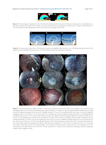
Figure 6. The pocket-creation method (PCM) of endoscopic submucosal dissection (ESD) for a locally recurrent tumor in the
rectosigmoid portion. (A) A laterally spreading tumor approximately 8 cm in diameter. (B) The small-caliber-tip transparent (ST) hood
entered the submucosal space below the tumor. Although using an ST hood with a small orifice, the entire view is secured through the
transparent wall of the ST hood. A focal submucosal scar resulting from one of multiple biopsies performed at a local hospital was
identified in the pocket. (C) The white surface of the muscularis was clearly identified distinguishing the blue submucosal layer in the
pocket. (D) Perforating blood vessels were identified very clearly in the pocket so that they were precisely coagulated without
immediate bleeding. (E) There was severe submucosal fibrosis due to previous multiple piecemeal endoscopic mucosal resections in
the central area of the tumor. (F) After dissecting the fibrosis, the tip of the endoscope reached a less fibrotic area beyond the fibrosis.
The submucosal pocket was subsequently opened. The ESD was completed in 211 min without any adverse events. (G) A mucosal
defect after the ESD did not have any thermal damage. (H) The back of the ESD specimen showed a white severe fibrotic area. (I) The
resected specimen and the tumor were 105 mm × 87 mm, and 82 mm × 75 mm in size, respectively. Pathology revealed a high-grade
adenoma with a negative margin.