
Page 11 - Read Online
P. 11
Ishihara. Mini-invasive Surg 2021;5:36 https://dx.doi.org/10.20517/2574-1225.2021.72 Page 5 of 9
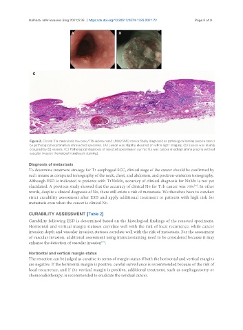
Figure 2. Clinical T1a-muscularis mucosae/T1b-submucosa 1 (MM/SM1) cancer finally diagnosed as pathological lamina propria cancer
by pathological examination of resected specimen. (A) Lesion was slightly elevated on white light imaging. (B) Lesion was mainly
occupied by B2 vessels. (C) Pathological diagnosis of resected specimen in our facility was cancer invading lamina propria without
vascular invasion (hematoxylin and eosin staining).
Diagnosis of metastasis
To determine treatment strategy for T1 esophageal SCC, clinical stage of the cancer should be confirmed by
such means as computed tomography of the neck, chest, and abdomen, and positron-emission tomography.
Although ESD is indicated to patients with T1N0M0, accuracy of clinical diagnosis for N0M0 is not yet
elucidated. A previous study showed that the accuracy of clinical N0 for T1b cancer was 73% . In other
[33]
words, despite a clinical diagnosis of N0, there still exists a risk of metastasis. We therefore have to conduct
strict curability assessment after ESD and apply additional treatment to patients with high risk for
metastasis even when the cancer is clinical N0.
CURABILITY ASSESSMENT [Table 2]
Curability following ESD is determined based on the histological findings of the resected specimens.
Horizontal and vertical margin statuses correlate well with the risk of local recurrence, while cancer
invasion depth and vascular invasion statuses correlate well with the risk of metastasis. For the assessment
of vascular invasion, additional assessment using immunostaining need to be considered because it may
enhance the detection of vascular invasion .
[34]
Horizontal and vertical margin status
The resection can be judged as curative in terms of margin status if both the horizontal and vertical margins
are negative. If the horizontal margin is positive, careful surveillance is recommended because of the risk of
local recurrence, and if the vertical margin is positive, additional treatment, such as esophagectomy or
chemoradiotherapy, is recommended to eradicate the residual cancer.