
Page 44 - Read Online
P. 44
Stier et al. Mini-invasive Surg 2020;4:18 I http://dx.doi.org/10.20517/2574-1225.2019.75 Page 5 of 11
Table 2. Results and comparison of upper endoscopy and 3D-CT
RYGB UE RYGB 3D-CT SG UE SG 3D-CT
Hiatal hernia Longitudinal measure (cm) 2.55 ± 0.82 2.24 ± 1.13 3.04 ± 1.23 2.69 ± 1.59
Volume (mL) 47.91 ± 20.86 174.41 ± 59.36
Diameter of the pouchoutlet (cm) 3.91 ± 0.71 2.16 ± 0.67
UE: upper endoscopy; 3D-CT: three-dimensional computed tomography volumetry; RYGB: roux-en-Y gastric bypass; SG: sleeve
gastrectomy
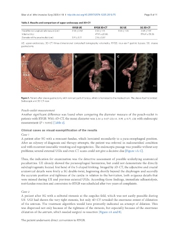
A B C
Figure 1. Patient after sleeve gastrectomy with remnant part of fundus, which is herniated to the mediastinum. The sleeve itself is twisted:
Endoscopic and 3D-CT view
Pouch-outlet measurement
Another significant difference was found when comparing the diameter measure of the pouch-outlet in
patients with RYGB. With 3D-CT, the mean diameter was 2.16 ± 0.67 cm vs. 3.91 ± 0.71 cm with endoscopic
measurement (P < 0.001) [Table 2].
Clinical cases as visual exemplification of the results
Case 1
A patient after SG with a remnant fundus, which herniated secondarily to a para-oesophageal position.
After an odyssey of diagnosis and therapy attempts, the patient was referred in malnourished condition
and with recurrent insatiable vomiting and regurgitation. The endoscopic passage was possible without any
problems; several external UGIs and even CT scans could not give a decisive clue [Figure 1A-C].
Thus, the indication for examination was the detective assessment of possible underlying anatomical
peculiarities. UE already showed the paraesophageal herniation, but could not demonstrate the directly
subdiaphragmatic located first bend of the S-shaped kinking. Imaged by 3D-CT, the adjunctive and crucial
anatomical details were firstly a SG double-twist, beginning shortly beyond the diaphragm and secondly
the accurate position and tightness of the cardia in relation to the herniation, both exiguous details that
were missed during UE and previous external UGIs. According those findings, immediate adhaesiolysis,
rest-fundus resection and conversion to RYGB was scheduled after two years of complaints.
Case 2
A patient after SG with a subtotal stenosis at the angulus fold, which was not easily passable during
UE. UGI had shown the very tight stenosis, but only 3D-CT revealed the enormous extent of dilatation
of the antrum. The treatment algorithm would have primarily indicated an attempt of dilation. This
was dispensed not only because of the tightness of the stenosis, but especially because of the enormous
dilatation of the antrum, which needed surgical re-resection [Figure 2A and B].
The patient underwent direct conversion to RYGB.