
Page 60 - Read Online
P. 60
Page 4 of 10 Mazzola et al. Mini-invasive Surg 2019;3:12 I http://dx.doi.org/10.20517/2574-1225.2019.05
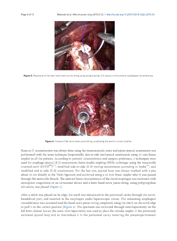
Figure 3. Placement of the hemi hand-sewn purse-string, using polypropylene 2/0 suture, on the anterior esophageal circumference
Figure 4. Closure of the hand-sewn purse-string, positioning the anvil in correct position
Roux-en-Y reconstruction was always done using the transmesocolic route and jejuno-jejunal anastomosis was
perforemed with the same technique (isoperistaltic side-to-side mechanical anastomosis using 45 mm linear
stapler) in all the patients. According to patients’ characteristics and surgeon preference, 3 techniques were
used for esophago-jejunal (E-J) anastomosis: hemi-double-stapling (HDS) technique using the transorally
[12]
TM [11]
inserted anvil (OrVil ) , modified side-to-side (S-S) overlap anastomosis according to Inaba , and
modified end-to-side (E-S) anastomosis. For the last one, jejunal loop was always marked with a pen
about 20 cm distally to the Treitz ligament and sectioned using a 45 mm linear stapler after it was passed
through the mesocolic breach. The anterior hemi-circumference of the distal esophagus was sectioned with
monopolar coagulation or an ultrasound device and a hemi hand-sewn purse-string, using polypropylene
2/0 suture, was placed [Figure 3].
After a stitch was placed on its edge, the anvil was introduced in the peritoneal cavity through the suvra-
humbilical port, and inserted in the esophagus under laparoscopic vision. The remaining esophageal
circumference was sectioned and the hand-sewn purse-string completed, using the stitch on the anvil edge
to pull it in the correct position [Figure 4]. The specimen was extracted through mini-laparotomy on the
left hemi-clavear trocar; the same mini-laparotomy was used to place the circular stapler in the previously
sectioned jejunal loop and to reintroduce it in the peritoneal cavity restoring the pneumoperitoneum