
Page 68 - Read Online
P. 68
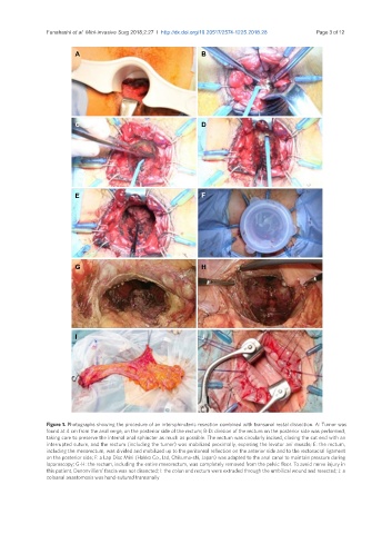
Funahashi et al. Mini-invasive Surg 2018;2:27  I  http://dx.doi.org/10.20517/2574-1225.2018.28                                  Page 3 of 12


                        A                                     B


                        C                                     D


                        E                                     F


                        G                                     H


                        I                                     J


               Figure 1. Photographs showing the procedure of an intersphincteric resection combined with transanal rectal dissection. A: Tumor was
               found at 4 cm from the anal verge, on the posterior side of the rectum; B-D: division of the rectum on the posterior side was performed,
               taking care to preserve the internal anal sphincter as much as possible. The rectum was circularly incised, closing the cut end with an
               interrupted suture, and the rectum (including the tumor) was mobilized proximally, exposing the levator ani muscle; E: the rectum,
               including the mesorectum, was divided and mobilized up to the peritoneal reflection on the anterior side and to the rectosacral ligament
               on the posterior side; F: a Lap Disc Mini (Hakko Co., Ltd, Chikuma-shi, Japan) was adapted to the anal canal to maintain pressure during
               laparoscopy; G-H: the rectum, including the entire mesorectum, was completely removed from the pelvic floor. To avoid nerve injury in
               this patient, Denonvilliers’ fascia was not dissected; I: the colon and rectum were extruded through the umbilical wound and resected; J: a
               coloanal anastomosis was hand-sutured transanally