
Page 215 - Read Online
P. 215
Page 4 of 13 Climent et al. Mini-invasive Surg 2018;2:45 I http://dx.doi.org/10.20517/2574-1225.2018.62
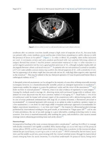
Figure 1. Anterior defect on end-to-end stapled anastomosis with a faecal collection
syndrome after an anterior resection should prompt a high index of suspicion of an AL, but some leaks
can present with a more insidious course and become evident later, presenting as a pelvic abscess or with
[16]
the presence of faeces in the pelvis [Figure 1]. In order to detect AL promptly, some earlier indicators
are used. A systematic review and meta-analysis performed with 2483 patients following colorectal
surgery showed that serum C-reactive protein concentration measured on day 3-5 after resection is a
useful negative predictive test but not a good positive predictor of AL, although included studies did not
[26]
distinguish between colonic and rectal resection . In patients who are not systemically unwell or unstable,
a CT with water-soluble contrast enema may confirm the clinical suspicion of AL, with a sensitivity of 0.91,
but the appearance of an intact staple line does not rule out an AL, with high false-negative rates reported
[27]
in the literature . This may be related to the fact that post-operative CT may be performed before there is
radiological evidence of AL .
[17]
A disrupted colorectal anastomosis can be salvaged in the majority of cases often utilising minimally invasive
[8]
techniques; however, in a haemodynamically unstable patient an emergent laparotomy is mandatory .
Laparoscopy enables the surgeon to assess the peritoneal cavity and the status of the anastomosis [16,28] with
[30]
[29]
faster recovery in selected patients . However, there is no clear evidence of superiority to open surgery .
Among the methods used to manage AL, diverting stoma with peritoneal lavage with or without intra-
[30]
abdominal drain placement was the most common method of managing AL . Small leaks (< 30% of the
circumference) could be treated with primary repair of the anastomosis with a defunctioning stoma, while
in case of severe peritoneal contamination with large AL or colonic ischaemia, a Hartmann’s procedure is
recommended . A transanal approach with anoscopy is an option in order to perform a primary repair on
[8]
a low anastomosis (< 5 cm from the anal verge) while a transanal endoscopic approach is recommended for
[28]
higher anastomosis (anastomosis ≥ 5 cm from anal verge) . The transrectal ultrasound-guided drainage
[16]
performed by interventional radiologists has good results, as mentioned in “Organ space SSI” and in case
of sinus persistence, the use of an endo-sponge is a good alternative, avoiding protecting stoma in some
scenarios. This device is inserted transanally, after washing the cavity, and attached to a low vacuum wound
[31]
drainage system enhancing granulating tissue and cavity closure .
Haemorrhage
[1]
Intraoperative bleeding is the most common intraoperative complication , and may be difficult to manage
with fatality imminent if not rapidly controlled. Pelvic haemorrhage can occur with injury of the presacral
venous plexus (PSVP) and the sacral basivertebral veins if dissection is posterior to the mesorectal plane,
behind the pre-sacral fascia, occurring in 4.6% to 9.4% of cases . PSVP is formed by the two lateral sacral
[32]
veins and the middle sacral vein, anastomosing with the internal vertebral system through the basivertebral
vessels emerging from the sacral foramina. It is localised underneath the presacral fascia, being easily