
Page 142 - Read Online
P. 142
Ambe et al. Mini-invasive Surg 2018;2:37 I http://dx.doi.org/10.20517/2574-1225.2018.45 Page 7 of 13
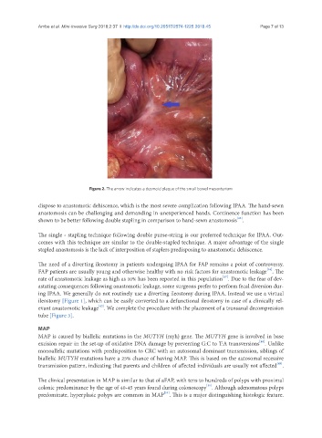
Figure 2. The arrow indicates a desmoid plaque of the small bowel mesenterium
dispose to anastomotic dehiscence, which is the most severe complication following IPAA. The hand-sewn
anastomosis can be challenging and demanding in unexperienced hands. Continence function has been
[45]
shown to be better following double stapling in comparison to hand-sewn anastomosis .
The single - stapling technique following double purse-string is our preferred technique for IPAA. Out-
comes with this technique are similar to the double-stapled technique. A major advantage of the single
stapled anastomosis is the lack of interposition of staplers predisposing to anastomotic dehiscence.
The need of a diverting ileostomy in patients undergoing IPAA for FAP remains a point of controversy.
[46]
FAP patients are usually young and otherwise healthy with no risk factors for anastomotic leakage . The
[47]
rate of anastomotic leakage as high as 10% has been reported in this population . Due to the fear of dev-
astating consequences following anastomotic leakage, some surgeons prefer to perform fecal diversion dur-
ing IPAA. We generally do not routinely use a diverting ileostomy during IPAA. Instead we use a virtual
ileostomy [Figure 1], which can be easily converted to a defunctional ileostomy in case of a clinically rel-
[42]
evant anastomotic leakage . We complete the procedure with the placement of a transanal decompression
tube [Figure 3].
MAP
MAP is caused by biallelic mutations in the MUTYH (myh) gene. The MUTYH gene is involved in base
[48]
excision repair in the set-up of oxidative DNA damage by preventing G:C to T:A transversions . Unlike
monoallelic mutations with predisposition to CRC with an autosomal dominant transmission, siblings of
biallelic MUTYH mutations have a 25% chance of having MAP. This is based on the autosomal recessive
[49]
transmission pattern, indicating that parents and children of affected individuals are usually not affected .
The clinical presentation in MAP is similar to that of aFAP, with tens to hundreds of polyps with proximal
[50]
colonic predominance by the age of 40-45 years found during colonoscopy . Although adenomatous polyps
[51]
predominate, hyperplasic polyps are common in MAP . This is a major distinguishing histologic feature.