
Page 430 - Read Online
P. 430
Panfoli et al. J Cancer Metastasis Treat 2020;6:35 I http://dx.doi.org/10.20517/2394-4722.2020.50 Page 5 of 8
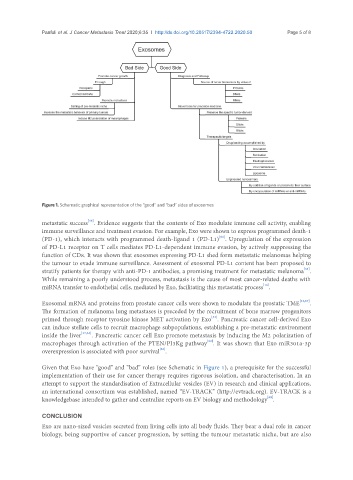
Figure 1. Schematic graphical representation of the “good” and “bad” sides of exosomes
[16]
metastatic success . Evidence suggests that the contents of Exo modulate immune cell activity, enabling
immune surveillance and treatment evasion. For example, Exo were shown to express programmed death-1
[61]
(PD-1), which interacts with programmed death-ligand 1 (PD-L1) . Upregulation of the expression
of PD-L1 receptor on T cells mediates PD-L1-dependent immune evasion, by actively suppressing the
function of CD8. It was shown that exosomes expressing PD-L1 shed form metastatic melanomas helping
the tumour to evade immune surveillance. Assessment of exosomal PD-L1 content has been proposed to
[61]
stratify patients for therapy with anti-PD-1 antibodies, a promising treatment for metastatic melanoma .
While remaining a poorly understood process, metastasis is the cause of most cancer-related deaths with
[16]
miRNA transfer to endothelial cells, mediated by Exo, facilitating this metastatic process .
Exosomal mRNA and proteins from prostate cancer cells were shown to modulate the prostatic TME [21,67] .
The formation of melanoma lung metastases is preceded by the recruitment of bone marrow progenitors
[33]
primed through receptor tyrosine kinase MET activation by Exo . Pancreatic cancer cell-derived Exo
can induce stellate cells to recruit macrophage subpopulations, establishing a pre-metastatic environment
inside the liver [17,33] . Pancreatic cancer cell Exo promote metastasis by inducing the M2 polarization of
[52]
macrophages through activation of the PTEN/PI3Kg pathway . It was shown that Exo miR301a-3p
[52]
overexpression is associated with poor survival .
Given that Exo have “good” and “bad” roles (see Schematic in Figure 1), a prerequisite for the successful
implementation of their use for cancer therapy requires rigorous isolation, and characterisation. In an
attempt to support the standardisation of Extracellular vesicles (EV) in research and clinical applications,
an international consortium was established, named “EV-TRACK” (http://evtrack.org). EV-TRACK is a
[68]
knowledgebase intended to gather and centralize reports on EV biology and methodology .
CONCLUSION
Exo are nano-sized vesicles secreted from living cells into all body fluids. They bear a dual role in cancer
biology, being supportive of cancer progression, by setting the tumour metastatic niche, but are also