
Page 338 - Read Online
P. 338
Page 16 of 31 Paul J Cancer Metastasis Treat 2020;6:29 I http://dx.doi.org/10.20517/2394-4722.2020.63
elevated glutaminolytic flux and enhanced amino acid and lipid metabolism. Some types of cancer cells
utilize in excess glucose and, in some cases secrete lactate even in the presence of oxygen (the Warburg
phenomenon). The propensity of cancer cells towards aerobic glycolysis does not seem to be related to an
impairment of the respiration, as respiration is also needed for tumor growth [130,131] . In some cancer patients,
lactate is converted back to glucose in the liver, a process known as the oncogenic Cori cycle [132-134] a process
that is energetically very inefficient. Besides glucose and lactate, there are other nutriments needed for
tumor growth for example, glutamine, glycine and aspartate for purine and pyrimidine synthesis, serine for
[135]
membrane lipid component synthesis, branched aminoacids, lipids, acetate and others . Not in all cancers
the Warburg phenomenon is present, and, sometimes, high glycolytic rates in tumors and mitochondrial
respiration often operate simultaneously in tumors [136] . A sort of metabolic parasitism has been described at
the tissular level by a group of French researchers [137] who introduced the concept of the “reverse Warburg
effect” [138,139] . These authors proposed that aggressive cancer cells are “parasites” that use oxidative stress
as a “weapon” to extract nutrients from surrounding stromal cells, forced to undergo aerobic glycolysis,
and produce energy-rich nutrients (such as lactate and ketones) to “feed” cancer cells. They suggested that
stromal catabolism, via autophagy and mitophagy, fuels the anabolic growth of tumor cells, promoting
tumor progression and metastasis.
What is also becoming apparent, is that cancer cells or tissues have an altered metabolism, but, they also
induce systemic changes of the whole body metabolism by secreting humoral factors (i.e., TNF‐α, IL‐1 and
IL‐6) and pro‐cachectic factors (i.e., proteolysis‐inducing factor and lipid mobilization factor) that lead to
a generalized catabolic state followed by significant and progressive energy loss from host tissue in the final
stages of cancer [140,141] . A group of researchers from Taiwan metaphorically compared these influences of
the tumor on the host’s metabolism as a “metabolic dictatorship”, the tumors imposing their high demands
on the normal host these metabolic changes, ultimately, in some types of cancers (i.e., pancreatic or gastric
cancer) leading to cachexia [142] . Basically, the metabolic parasitism described at the tissular level exists also
at the level of the whole organism [143] .
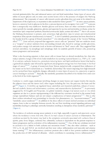
Cachexia is a multi-organ syndrome involving changes in many tissues and organs besides the muscle,
the adipose tissue and the tumor itself, other organs including the liver, the pancreas, the brain and the
gut [144] [Figure 8]. It involves up‐regulated tissue catabolism and impaired anabolism, release of tumor‐
derived catabolic factors and inflammatory cytokines, and neuroendocrine dysfunction [145] . As previously
suggested by Al-Zoughbi and Porporato, the global metabolic changes that tumors exert on the whole
organism are due to a precise reprogramming of the different key structures involved in the normal body
energy expenditure balance and are not simply complications of tumor progression [6,134] . This is why
[6]
these authors introduced new terminology to describe this phenomenon, i.e., “macroenvironment” and
“metabolic cancer syndrome” [134] . In addition to the direct effects of tumor-derived cytokines on individual
organs, there is also an interplay between muscle, fat, and liver involving several signaling pathways and
metabolites leading to pathologic networks formation resulting in disruption of key metabolic pathways [140]
[Figure 8].
The incidence of cachexia among cancer patients is very high, especially in gastric and pancreatic cancer
where the incidence is more than 80%. One of the main causes of cancer cachexia is inflammation. The
cytokines secreted by the tumor may lead to the symptoms commonly associated with cachexia (loss of
apetite, pain, fever, fatigue, cachexia) but, ultimately, cachexia is dependent on the patient response to
tumor progression and the activation of the inflammatory response [146] . One of the key cytokines involved
[86]
in cachexia is IL-6, linked to both cachexia and metastasis events , but, also other cytokines - such as
TNF‐α, IL-1β, and TGF-β- are involved and they may induce inflammation and muscular and adipose
tissue wasting [147,148] . Pro-inflammatory cytokines promote also a shift in liver protein synthesis towards
the production of CRP instead of albumin - which contributes to sustaining chronic inflammation [149] .