
Page 180 - Read Online
P. 180
Page 4 of 9 Lo Re et al. J Cancer Metastasis Treat 2020;6:17 I http://dx.doi.org/10.20517/2394-4722.2020.11
A B
C D
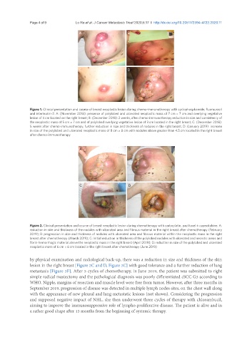
Figure 1. Clinical presentation and course of breast neoplastic lesion during chemo-immunotherapy with cyclophosphamide, fluorouracil
and interleukin-2. A: (November 2018): presence of polylobed and ulcerated neoplastic mass of 7 cm × 7 cm and overlying vegetative
lesion of 4 cm located on the right breast; B: (December 2018): 2 weeks, after chemo-immunotherapy reduction in size and consistency of
the neoplastic mass of 5 cm × 7 cm and of polylobed overlying vegetative lesion of 3 cm located in the right breast; C: (December 2018):
5 weeks after chemo-immunotherapy, further reduction in size and thickness of nodules in the right breast; D: (January 2019): increase
in size of the polylobed and ulcerated neoplastic mass of 8 cm × 8 cm with nodules above greater than 4.5 cm located in the right breast
after chemo-immunotherapy
A B
C D
Figure 2. Clinical presentation and course of breast neoplastic lesion during chemotherapy with carboplatin, paclitaxel ± capecitabine. A:
reduction in size and thickness of the nodules with ulcerated area and fibrous material in the right breast after chemotherapy (February
2019); B: progression in size and thickness of nodules with ulcerated area and fibrous material within the neoplastic mass in the right
breast after chemotherapy (March 2019); C: initial reduction in thickness of the polylobed nodules with ulcerated and necrotic areas and
fibrin-hemorrhagic material above the neoplastic mass in the right breast (April 2019); D: reduction in size of the polylobed and ulcerated
neoplastic mass of 6 cm × 6 cm located in the right breast after chemotherapy (June 2019)
by physical examination and radiological back-up, there was a reduction in size and thickness of the skin
lesion in the right breast [Figure 2C and D, Figure 3C] with good tolerance and a further reduction of lung
metastasis [Figure 3F]. After 5 cycles of chemotherapy, in June 2019, the patient was submitted to right
simple radical mastectomy and the pathological diagnosis was poorly differentiated cSCC G3 according to
WHO. Nipple, margins of resection and muscle level were free from tumor. However, after three months in
September 2019, progression of disease was detected in multiple lymph nodes sites, on the chest wall along
with the appearance of new pleural and lung metastatic lesions (not shown). Considering the progression
and supposed negative impact of NHL, she then underwent three cycles of therapy with chlorambucil,
aiming to improve the immunosuppressive role of lympho-proliferative disease. The patient is alive and in
a rather good shape after 13 months from the beginning of systemic therapy.