
Page 127 - Read Online
P. 127
Miliotis et al. J Cancer Metastasis Treat 2020;6:13 I http://dx.doi.org/10.20517/2394-4722.2020.12 Page 3 of 15
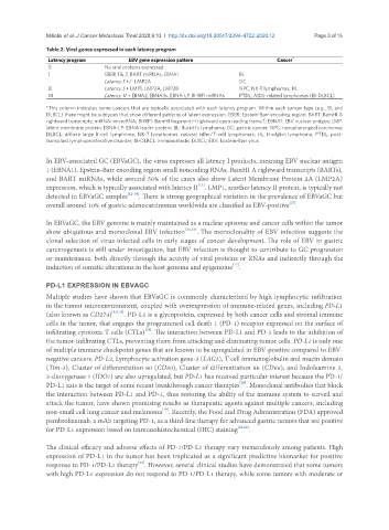
Table 2. Viral genes expressed in each latency program
Latency program EBV gene expression pattern Cancer *
0 No viral proteins expressed
I EBER 1 & 2, BART miRNAs, EBNA1 BL
Latency I +/- LMP2A GC
II Latency I + LMP1, LMP2A, LMP2B NPC, NK-T lymphomas, HL
III Latency II + EBNA2, EBNA3s, EBNA-LP, BHRF1 miRNAs PTDL, AIDS-related lymphomas (IB-DLBCL)
*This column indicates some cancers that are typically associated with each latency program. Within each cancer type (e.g., BL and
DLBCL) there might be subtypes that show different patterns of latent expression. EBER: Epstein-Barr encoding region; BART: BamHI A
rightward transcripts; miRNA: microRNA; BHRF1: BamHI fragment H rightward open reading frame 1; EBNA1: EBV nuclear antigen; LMP:
latent membrane protein; EBNA-LP: EBNA leader protein; BL: Burkitt’s lymphoma; GC: gastric cancer; NPC: nasopharyngeal carcinoma;
DLBCL: diffuse large B-cell lymphoma; NK-T lymphomas: natural killer/T-cell lymphomas; HL: Hodgkin lymphoma; PTDL: post-
transplant lymphoproliferative disorder; IB-DLBCL: immunoblastic DLBCL; EBV: Epstein-Barr virus
In EBV-associated GC (EBVaGC), the virus expresses all latency I products, meaning EBV nuclear antigen
1 (EBNA1), Epstein-Barr encoding region small noncoding RNAs, BamHI A rightward transcripts (BARTs),
and BART miRNAs, while around 50% of the cases also show Latent Membrane Protein 2A (LMP2A)
[12]
expression, which is typically associated with latency II . LMP1, another latency II protein, is typically not
detected in EBVaGC samples [12-14] . There is strong geographical variation in the prevalence of EBVaGC but
[15]
overall around 10% of gastric adenocarcinomas worldwide are classified as EBV-positive .
In EBVaGC, the EBV genome is mainly maintained as a nuclear episome and cancer cells within the tumor
show ubiquitous and monoclonal EBV infection [12,16] . The monoclonality of EBV infection suggests the
clonal selection of virus-infected cells in early stages of cancer development. The role of EBV in gastric
carcinogenesis is still under investigation, but EBV infection is thought to contribute to GC progression
or maintenance, both directly through the activity of viral proteins or RNAs and indirectly through the
[17]
induction of somatic alterations in the host genome and epigenome .
PD-L1 EXPRESSION IN EBVAGC
Multiple studies have shown that EBVaGC is commonly characterized by high lymphocytic infiltration
in the tumor microenvironment, coupled with overexpression of immune-related genes, including PD-L1
(also known as CD274) [4,5,18] . PD-L1 is a glycoprotein, expressed by both cancer cells and stromal immune
cells in the tumor, that engages the programmed cell death 1 (PD-1) receptor expressed on the surface of
[19]
infiltrating cytotoxic T cells (CTLs) . The interaction between PD-L1 and PD-1 leads to the inhibition of
the tumor-infiltrating CTLs, preventing them from attacking and eliminating tumor cells. PD-L1 is only one
of multiple immune checkpoint genes that are known to be upregulated in EBV-positive compared to EBV-
negative cancers. PD-L2, Lymphocyte activation gene-3 (LAG3), T cell immunoglobulin and mucin domain
(Tim-3), Cluster of differentiation 80 (CD80), Cluster of differentiation 86 (CD86), and Indoleamine 2,
3-dioxygenase 1 (IDO1) are also upregulated, but PD-L1 has received particular interest because the PD-1/
[20]
PD-L1 axis is the target of some recent breakthrough cancer therapies . Monoclonal antibodies that block
the interaction between PD-L1 and PD-1, thus restoring the ability of the immune system to surveil and
attack the tumor, have shown promising results as therapeutic agents against multiple cancers, including
[19]
non-small cell lung cancer and melanoma . Recently, the Food and Drug Administration (FDA) approved
pembrolizumab, a mAb targeting PD-1, as a third-line therapy for advanced gastric tumors that are positive
for PD-L1 expression based on immunohistochemical (IHC) staining [21,22] .
The clinical efficacy and adverse effects of PD-1/PD-L1 therapy vary tremendously among patients. High
expression of PD-L1 in the tumor has been implicated as a significant predictive biomarker for positive
[23]
response to PD-1/PD-L1 therapy . However, several clinical studies have demonstrated that some tumors
with high PD-L1 expression do not respond to PD-1/PD-L1 therapy, while some tumors with moderate or