
Page 100 - Read Online
P. 100
Page 4 of 9 Ray et al. J Cancer Metastasis Treat 2020;6:9 I http://dx.doi.org/10.20517/2394-4722.2020.16
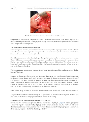
Figure 3. Diaphragmatic rent after partial diaphragmatic excision (black arrow)
not performed. We aspirated the pleural effusion in each case and dissected a few pleural deposits with
electro-diathermy in one case. Thorough pleural lavage with chemotherapeutic perfusate into the pleural
cavity was performed during HIPEC.
The technique of diaphragmatic resection
For diaphragmatic resection, one must be aware of the anatomy of the diaphragm in relation to the phrenic
nerve. The phrenic nerve originates mainly from the 4th cervical nerve, but also receives contributions
from the 5th and 3rd cervical nerves (C3-C5).
The right phrenic nerve enters the diaphragm through the central tendon or inferior vena cava opening.
On the right side, it courses relatively more medially throughout its thoracic course to various structures
like the right brachiocephalic vein, SVC and pericardium over the right atrium. The inferior vena cava
lies medially and reaches under the surface of the diaphragm by passing through the inferior vena cava
foramen in the central tendon.
The left phrenic nerve pierces the superior surface of the muscular part of the diaphragm, just to the left
border of the heart.
Both nerves divide or trifurcate at, or just above the diaphragm. The branches travel together into the
diaphragmatic musculature, while small sensory branches supply the peritoneum over the central part of
the diaphragm. The larger motor branches separate within the diaphragm into four major nerves trunks -
sternal, anterolateral, postero-lateral and crural. The nerve trunks travel partly within the diaphragmatic
muscle and innervate the inferior surface covered by peritoneum. Therefore, the diaphragmatic incision
has to be made circumferentially to avoid the main phrenic nerve trunks.
In the present study, we made an incision in the above-mentioned manner and excised the tumor deposits.
The patient’s head end was lowered during HITAC procedures, so that the chemotherapeutic fluid can gain
easy access to the thoracic cavity by free flow from the abdominal cavity.
Reconstruction of the diaphragm after HITAC procedure
In two cases, we had to incise approximately one-fourth of the diaphragm [Figure 3]. The diaphragmatic
defects were repaired primarily with polypropylene 1-0 suture. In another case, the peritoneal defect was
almost 50% and required reconstruction with polypropylene mesh, fixed with polypropylene 1-0 suture. In
all three cases, we placed the ICD in the triangle of safety.