
Page 816 - Read Online
P. 816
Fiordoliva et al. J Cancer Metastasis Treat 2019;5:59 I http://dx.doi.org/10.20517/2394-4722.2019.23 Page 3 of 8
A B
C D
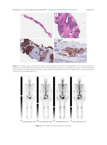
Figure 1. A: Prostate biopsy extensively involved by acinar adenocarcinoma with NE differentiation (5%) at low magnification
(hematoxylin and eosin-H&E); B: prostate biopsy with solid area of adenocarcinoma at high magnification (H&E); C: prostate biopsy with
diffuse positivity for prostate specific membrane antigen (PSMA, immunohistochemistry); D: prostate biopsy with focal positivity for
synaptophysin (immunohistochemistry)
Figure 2. Bone scintigraphy confirming skeletal involvement