Page 478 - Read Online
P. 478
Bookland et al. J Cancer Metastasis Treat 2019;5:33  I  http://dx.doi.org/10.20517/2394-4722.2018.110                      Page 5 of 16

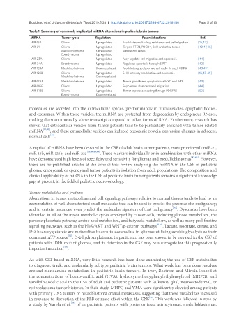
               Table 1. Summary of commonly implicated miRNA alterations in pediatric brain tumors
                MiRNA          Tumor types     Regulation             Potential actions           Ref.
                MiR-15B        Glioma           Upregulated  Modulates multi-drug resistance and cell migration  [16,47]
                MiR-21         Glioma           Upregulated  Targets PTEN, PDCD4, Bcl2 and other tumor   [42,47,48]
                               Medulloblastoma  Upregulated  suppressor genes.
                               Ependymoma       Upregulated
                MiR-23A        Glioma           Upregulated  May regulate cell migration and apoptosis.  [44]
                MiR-34A        Ependymoma       Upregulated  Regulates apoptosis through SiRT1   [42]
                MiR-124A       Medulloblastoma  Downregulated  Modulates glycolysis and cell cycle through CDK6  [43,49]
                MiR-125B       Glioma           Upregulated  SHH pathway modulation and apoptosis  [16,47-49]
                               Medulloblastoma  Downregulated
                MiR-128A       Medulloblastoma  Upregulated  Tumor growth and apoptosis via MYC and Bcl2  [43]
                MiR-146B       Glioma           Upregulated  Suppresses stemness and migration   [44]
                MiR-518B       Glioma           Upregulated  Tumor suppressor acting through PDGFRB  [50]
                               Ependymoma       Downregulated


               molecules are secreted into the extracellular spaces, predominantly in microvesicles, apoptotic bodies,
               and exosomes. Within these vesicles, the miRNA are protected from degradation by endogenous RNases,
               making them an unusually stable transcript compared to other forms of RNA. Furthermore, research has
               shown that extracellular vesicles from tumor patients tend to be particularly enriched with tumor-related
               miRNA  [44,45] , and these extracellular vesicles can induced oncogenic protein expression changes in adjacent,
               normal cells .
                         [46]
               A myriad of miRNA have been detected in the CSF of adult brain tumor patients, most prominently miR-21,
               miR-15b, miR-125b, and miR-223 [16,44,48,49] . These markers individually or in combination with other miRNA
               have demonstrated high levels of specificity and sensitivity for gliomas and medulloblastomas [47,49] . However,
               there are no published articles at the time of this review analyzing the miRNA in the CSF of pediatric
               glioma, embryonal, or ependymal tumor patients in isolation from adult populations. The composition and
               clinical applicability of miRNA in the CSF of pediatric brain tumor patients remains a significant knowledge
               gap, at present, in the field of pediatric neuro-oncology.

               Tumor metabolites and proteins
               Aberrations in tumor metabolism and cell signaling pathways relative to normal tissues tends to lead to an
               accumulation of well-characterized small molecules that can be used to predict the presence of a malignancy;
                                                                                     [51]
               and in certain instances, even predict the molecular signature of that malignancy . Dyscrasias have been
               identified in all of the major metabolic cycles employed by cancer cells, including glucose metabolism, the
               pentose phosphate pathway, amino acid metabolism, and fatty acid metabolism, as well as many proliferative
               signaling pathways, such as the PI3K/AKT and WNT/β-catenin pathways [8,16] . Lactate, isocitrate, citrate, and
               D-2-hydroxyglutarate are metabolites known to accumulate in gliomas utilizing aerobic glycolysis as their
               dominant ATP source . D-2-hydroxyglutarate, in particular, has been shown to be elevated in the CSF of
                                  [52]
               patients with IDH1 mutant gliomas, and its detection in the CSF may be a surrogate for this prognostically
                                [53]
               important mutation .
               As with CSF-based miRNA, very little research has been done examining the use of CSF metabolites
               to diagnose, track, and molecularly subtype pediatric brain tumors. What work has been done revolves
               around monoamine metabolism in pediatric brain tumors. In 1987, Bostrom and Mirkin looked at
               the concentrations of homovanillic acid (HVA), hydroxymethoxyphenylethyleneglycol (MHPG), and
               vanillylmandelic acid in the CSF of adult and pediatric patients with leukemia, glial, neuroectodermal, or
               retinoblastoma tumor histories. In their study, MHPG and VMA were significantly elevated among patients
               with primary CNS tumors or neuroblastoma cranial metastases, suggesting that these metabolites increased
                                                                         [54]
               in response to disruption of the BBB or mass effect within the CNS . This work was followed in 2014 by
               a study by Varela et al.  of 22 pediatric patients with posterior fossa astrocytomas, medulloblastomas,
                                   [55]

