Page 392 - Read Online
P. 392
Bonfiglio et al. J Cancer Metastasis Treat 2019;5:29  I  http://dx.doi.org/10.20517/2394-4722.2018.88                          Page 5 of 7


                                                 P = 0.0030
                              Positive cells/500


                                                 P = 0.0024
                               Positive cells/500


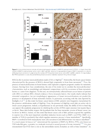
               Figure 2. Immunohistochemical analysis of bone morphogenetic proteins 2 (BMP-2) and pentraxin-3 (PTX3). A: Graph shows the
               number of BMP-2 positive breast cancer cells in BM+ and BM- patients; B: representative image of BMP-2 expression in a ductal
               infiltrating breast carcinoma; C: graph shows the number of PTX3 positive breast cancer cells in BM+ and BM- patients; D: image displays
               the PTX3 expression in a case of ductal infiltrating breast carcinoma. Scala bars represent 100 μm for all images

                                                                        [7]
               BOLCs) able to product microcalcifications made of HA or MgHAp . Noteworthy, the breast cancer lesions
                                                                                               [7]
               characterized by the presence of BOLCs showed high propensity to form bone metastasis . Thus, the
               presence of microcalcifications in breast lesions could represent a negative prognostic marker for metastatic
               diseases. Starting from these considerations, the aim of this study was to correlate the microcalcifications’
               characteristics, such as morphology and elemental compositions, with the occurrence of bone metastatic
               lesions at 5 years from diagnosis. To this end, we retrospectively collected breast cancer lesions of patients
               with (BM+) or without (BM-) clinical evidence of bone metastatic lesions. Our results clearly indicate that
               elemental composition, but not morphological appearance, of breast microcalcifications can predict the
               presence or development of bone metastatic lesions. In particular, according with the data reported by
                            [15]
               Bonfiglio et al. , in this study the breast cancer lesions of BM+ patients were frequently characterized by
               the presence calcifications made of MgHAp. Thus, the presence of MgHAp could play an active role in
               breast cancer progression. Indeed, as hypothesized in our previously study, the ability of HA to bind Mg
               can support the neoplastic progression by inhibiting the activity of DNA repair enzymes that require Mg as
               co-factor. These data further support the hypothesis that the bone metastatic process of breast cancer can
               be driven by BOLCs. Indeed, we also noted that breast cancer cells of BM+ patients acquired the capability
               to express two of the most important osteoblast induction factors such as BMP-2 and PTX3. BMP-2 is a
                                                                                             [8]
               member of TGF-β superfamily that which regulate numerous process in bone metabolisms . Specifically,
                                                                                                        [8]
               BMP-2 is involved in both mesenchymal stem cells recruitment and differentiation into mature osteoblasts .
                                          [7]
               In a recent paper, Scimeca et al.  demonstrated the expression of BMP-2 by BOLCs. PTX3, also known as
               TNF-inducible gene 14 protein, is a molecule involved in several process of innate immunity [17,18] . It is also
                                                                                                     [9]
               demonstrated that PTX3 have a role in the extracellular matrix formation as well as bone formation . In

